Health Library
Ventricular Septal Defect

What is a Ventricular Septal Defect (VSD)?

A ventricular septal defect (VSD) is a hole between the right and left pumping chambers of the heart.

The heart has four chambers: a right and left upper chamber called an atrium and a right and left lower chamber called a ventricle.

In the normal heart, the right and left chambers are separated from each other by a wall of muscle called a septum. The right atrium is separated from the left atrium by the atrial septum. The right ventricle is separated from the left ventricle by the ventricular septum.

It is normal for all infants to be born with a small hole between the two atria which usually closes within the first few weeks of life.

Normally there is no hole between the two ventricles, but some infants are born with these holes called ventricular septal defects.

Ventricular septal defects are among the most common congenital heart defects, occurring in 0.1 to 0.4 percent of all live births. Ventricular septal defects are one of the most common reasons for infants to see a cardiologist (a doctor who treats the heart).

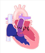
View 2D images of Ventricular Septal Defect.

Learn about the Heartpedia mobile app. The app shows anatomically accurate images of congenital heart defects and repairs of those defects.

Description of Ventricular Septal Defects

Ventricular septal defects occur in many locations and sizes. The ventricular septum is made up of different types of tissue, with one part composed of mainly muscle and another part made of thinner, fibrous tissue. The location and size of the hole within the septum will determine in part how to treat the ventricular septal defect.

Small ventricular septal defects rarely cause problems. A doctor usually discovers these holes by noticing an extra heart sound called a murmur, on a routine physical exam. This murmur is often not present in the first few days of life.

Most of these holes will close on their own, particularly if they are in the muscular portion of the septum. Even if these holes do not close, they will rarely cause any health problems.

These holes can connected to the development of other heart issues. If the small ventricular septal defect does not close, the child should continue to be seen by a cardiologist for occasional checkups.

Large ventricular septal defects can cause problems, often developing gradually in the first few months of life. Before birth, the pressure on the right side of the heart is equal to pressure on the left side of the heart.

As soon as the baby takes its first breath, the pressure in the lungs and the right side of the heart starts to decrease. This process is slow and usually takes about two to four weeks for the pressure in the lungs to reach normal level.

In the first one to two weeks of life, babies with large ventricular septal defects may do very well. As the pressure in the right side of the heart decreases, blood will start to flow to the path of least resistance (from the left ventricle through the ventricular septal defect to the right ventricle and into the lungs). This will gradually lead to symptoms of congestive heart failure and must be treated.

Medium or moderate ventricular septal defects are more challenging to predict. Sometimes babies born with moderate ventricular septal defects will have problems with congestive heart failure like babies with large ventricular septal defects. Others will have no problems and will need to be watched.

Ventricular septal defects never get bigger and sometimes get smaller or close completely. When a baby is diagnosed with a ventricular septal defect, most cardiologists will not recommend immediate surgery. They will closely observe the baby and try to treat symptoms of congestive heart failure with medicine to allow time to determine if the defect will close on its own.

Signs and Symptoms

Ventricular septal defects have a very characteristic murmur, to the point where a cardiologist may be able to pinpoint the location and estimate the size of a ventricular septal defect just by how it sounds.

A murmur is often not heard at birth. It is only with time and pressure changes that flow across the hole between the pumping chambers can be heard as a murmur.

A smaller hole may make a louder noise than a large hole, and the murmur may get louder as the ventricular septal defect closes.

Think of a garden hose. If the water flows freely, it makes a soft sound. If you make the outlet of the hose smaller with your finger, the noise will get louder. It is important to remember a loud murmur does not mean a large hole.

Babies who do have moderate or large ventricular septal defects with excessive blood flow to the lungs will have signs of congestive heart failure. The most important sign will be the baby's growth.

Babies who have significant congestive heart failure will have failure to thrive and will have difficulty maintaining a normal weight gain in the first few months of life.

Babies with some extra flow to the lungs may grow well because their ability to feed remains unaffected. They may have some subtle signs of congestive heart failure such as continuous fast breathing.

If a baby grows well in the first few months, it is likely that the ventricular septal defect will not lead to congestive heart failure. If the baby does show significant signs of congestive heart failure, the ventricular septal defect may need to be surgically closed.

Diagnosis of Ventricular Septal Defects

Most ventricular septal defects can be diagnosed on physical exam, due to their murmur. The murmur can change with time due to the hole closing, or due to more blood flow across the hole.

The heart can sometimes be seen or felt to be beating hard because of the extra work it is performing. Babies can be continuously breathing fast or hard and have a fast heart rate.

An electrocardiogram can help determine the sizes of the chambers to see if there is strain on the heart due to the ventricular septal defect. The electrocardiogram can be normal at birth and change with time as congestive heart failure worsens. It can also suggest if there are other heart defects associated with the ventricular septal defect.

A chest X-ray can help follow the progression of congestive heart failure by looking at the size of the heart and the amount of blood flow to the lungs. This may be normal at birth and change with time.

Most small ventricular septal defects will not require an echocardiogram as they tend to close, but often infants with moderate or large ventricular septal defects will need to have at least one echocardiogram to provide the cardiologist with a complete picture of the defect.

In some children with ventricular septal defects a cardiac catheterization will need to be performed. This can help the cardiologist determine how much blood flow is going out to the lungs. This can be useful in determining the need for surgery in children who have had subtle signs of congestive heart failure but who do not have clear-cut evidence of the need for surgical repair.

Treatment for Ventricular Septal Defects

Many times observation is the only treatment needed, with regular checkups with the cardiologist. This may be every two to three years in older children with small ventricular septal defects to as often as weekly in babies with large ventricular septal defects.

Babies who have shown some signs of congestive heart failure will typically be placed on medicine, usually a diuretic to help get rid of extra fluid in the lungs.

In babies who are failing to thrive because it is too difficult for them to eat, a high calorie formula or fortified breast milk will be added to help the baby grow.

Sometimes babies get worn out with feeding. A small tube may need to be inserted through the nose and into the stomach. This is temporary to help deliver the food. The goal is to control the symptoms of heart failure to allow the baby time to grow.

The ventricular septal defect may get smaller and cause fewer problems. The infant will not require surgery and will eventually not need medicines.

When the symptoms of a ventricular septal defect are hard to control with medicines or the baby is unable to grow, surgical closure of the defect is often recommended. Surgical closure of isolated ventricular septal defects is uncomplicated in 99 percent or more of cases.

Some ventricular septal defects may be closed using an FDA approved closure device which is placed using a heart catheter (a small plastic tube through which the device may be delivered). In infants this requires a more limited surgery to be used together with catheter placement of the device.

Long-Term Outlook

Small ventricular septal defects which do not eventually close rarely cause any longer-term difficulties. However, depending on the location of the hole, lifelong follow-up may be required.

Children who have had their ventricular septal defects close on their own or closed completely at surgery do not need any medicines, and should not be restricted in any way.

Unrepaired large ventricular septal defects and some moderate ventricular septal defects can cause two problems.

First, infants may have ongoing symptoms of congestive heart failure. Infants who have poor growth due to congestive heart failure can have poor brain development during the first few years of life. This is the time of most rapid brain development, therefore good nutrition is important for the development of the baby.

Babies with congestive heart failure are at higher risk for infections, particularly lung infections. If they do get a lung infection, they may not tolerate it as well as other babies and can become very sick with even a winter cold. Immunizations may protect against some lung infections but not all, and complications from these infections can be life-threatening.

Secondly, as the lungs are exposed to excessive pressure and flow over a period of years the vessels in the lungs may react by developing thicker walls. The pressures in the lungs will then increase as a result.

The pressures in the lungs can become so high that blue (blood with little oxygen) blood from the right ventricle will flow across the ventricular septal defect into the left ventricle and mix with red (blood with oxygen) blood.

The patient will then have less oxygen going to the rest of the body and will start to develop cyanosis (blue coloring of lips and nails). The high pressures in the lungs can initially be reversed, but with time will become irreversible and result in failure of the heart.

If large ventricular septal defects are diagnosed and managed appropriately, a child with a ventricular septal defect can have a normal length of life with no restrictions.

Last Updated 05/2022

Reviewed By Kathy Meyer, RN

Contact

Contact us.
A national leader in pediatric and congenital heart surgery and transplantation, we take on patients with complex conditions that other hospitals are unable to treat. Contact Us

Locations Close to Home

Learn more about visiting Cincinnati Children's.

The Heart Institute has more than 30 outpatient heart locations in Ohio, Kentucky and Indiana.

Find a Location