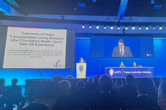
Cincinnati Children's Hospital Medical Center Logo

Eghtesady-Clark Lab

Research in the Lab

Research Images
A representative picture of an ovine fetal heart 1 month after banding.
Significant LVH with Aortic Banding.
Close up view of a 95 day sheep fetal (~26 weeks humans) heart prior to surgery. Anatomic landmarks are identified.
[Click for detailed image.]

A major focus of research in the lab is to define the mechanisms in the pathogenesis of Hypoplastic Left Heart Syndrome (HLHS), which has highest mortality rate among congenital heart defects. The primary feature of HLHS is a left ventricle that is too small or "hypoplastic" to support circulation after birth. It is believed that in utero aortic stenosis, a defect presenting as narrowing or obstruction of the fetal aortic valve, can lead to development of HLHS. The etiology of HLHS and the morphogenesis of left ventricular hypoplasia remain unknown. Details of HLHS can be found in the Heart Center Encyclopedia.

Below is a brief synopsis of these projects:

Development of an Animal Model of HLHS or Aortic Stenosis

Using the fetal sheep model, we have studied mechanisms of left heart hypoplasia such as would occur in fetal aortic stenosis. Progressive supravalvar aortic stenosis is induced in early-gestation sheep fetuses (equivalent to 11-17 weeks human gestation) by surgically placing a band around the fetal ascending aorta (top panel). The"in vivo" natural history of the induced changes is followed through serial fetal echocardiography. Simultaneously cardiac catheterization analysis is done to assess physiologic changes by alterations in blood flow in the fetal heart (middle panel). A substantial reduction in left ventricular chamber volume is induced in the banded (bottom panel - image on left) compared with the control heart (bottom panel - image on right).

Preliminary work suggests that pressure overload is not sufficient to cause HLHS.  Previous studies by other investigators have been conducted in the 3rd trimester fetal sheep.  Our research is the first to study the 1st trimester fetal sheep model. 

Brain Natriuretic Peptide as a Marker of Fetal Cardiovascular Health

Despite the prevalence of fetal heart disease, a limited amount of information exists upon which to characterize the cardiovascular health of a fetus. A key step in improving the effectiveness of fetal cardiac intervention will be the advent of a quantitative indicator of systemic fetal cardiovascular health to augment data obtained via fetal echocardiography. Our lab has created a reproducible model of fetal aortic stenosis in the early gestation sheep fetus via surgical manipulation of the fetal ascending aorta.  Our hypothesis is that amniotic fluid brain natriuretic peptide (BNP) concentrations are altered in cases of fetal cardiac malformations, and thus that amniotic BNP levels can serve a prognostic value for fetal cardiac health.

Fetal Therapy for Fetal Aortic Stenosis

(Collaborator: Dr. Eric C. Michelfelder, MD, Division of Cardiology and The Fetal Care Center of Cincinnati, Cincinnati Children's Hospital). This project is focused on identifying and developing optimal techniques of fetal cardiac intervention for a heart defect called aortic stenosis.

Characterized by a narrowing of the aortic valve (the main valve before blood leaves the heart to go to the body), aortic stenosis is believed to be the precursor for developing Hypoplastic Left Heart Syndrome by limiting blood flow through the left side of the heart and therefore, stunting its growth. Many are now attempting to relieve this narrowing with fetal intervention in the clinical setting (primarily at Boston Children's Hospital) but with limited success. The purpose of this project is test alternate techniques and to also develop methods for better selection of appropriate candidates for the procedures. This project was started in 2004 and involves animal experiments that are preliminary to clinical applications here at Cincinnati Children's Hospital. It is anticipated that better selection and improved techniques (discovered through this research) will result in higher success rate (currently ~15%) with fetal cardiac intervention. We anticipate approximately 1 to 2 years prior to clinical application of the research. A segment of the research, involving use of a protein marker in the amniotic fluid as a diagnostic and prognostic marker of severity of heart disease, has already been approved by Cincinnati Children's Hospital Institutional Review Board for clinical testing and a patent application is also pending through the Internal Patent office. 

Development of Techniques for Fetal Cardiac Surgery

(Collaborators: Drs. Kenneth Clark, PhD and Leslie Myatt, PhD, Department of Obstetrics and Gynecology, University of Cincinnati and Dr. Eric C. Michelfelder, MD, Division of Cardiology and The Fetal Care Center of Cincinnati, Cincinnati Children's Hospital). The focus of this project deals with development of tools and techniques for clinical application of fetal open-heart surgery, a requisite for management of certain fetal heart defects not amenable to other less-invasive treatment modalities. These would include heart defects that are either lethal prenatally or cause such severe damage to the heart during prenatal life that death is imminent once born (for example in babies with a variant of Hypoplastic Left Heart Syndrome, involving an intact, who have a mortality close to 100% despite "optimal" therapy).

Using the fetal sheep model, we have studied mechanisms of left heart hypoplasia such as would occur in fetal aortic stenosis. Progressive supravalvar aortic stenosis was induced in early-gestation sheep fetuses, equivalent to 11-17 weeks human gestation (Figure 1 A representative photograph of the fetal heart). The research is still at the "bench" level, but it is anticipated to be ready for preliminary testing within 3 years (a single clinical attempt has already been made at Stanford University Hospital without success by my prior mentor, Dr. Frank Hanley).

Our Perfusion Group (Robert E. Ferguson, CCP, Aimee B. Gardner, CCP, John P. Lombardi, CCP and Jerri L. McNamara, CCP) is critically involved in this project as much of this technology depends on the application of cardiopulmonary bypass during surgery.

Cardiopulmonary bypass technology permits open-heart surgery because it temporarily bypasses and functions for the heart and lungs. Other terms used for cardiopulmonary bypass include extracorporeal circulation and heart-lung machine (referring to the actual bypass machinery). Although this technology is quite sophisticated, the concept is rather simple: blood is removed from the heart through surgically placed venous cannulas and delivered to a heart-lung machine that oxygenates, heats and filters the blood (can also add drugs) before pumping it back to the fetus through another surgically placed arterial cannula. While no one has successfully applied this technology to a human fetus, it has been done with limited success in the experimental setting.
 
To carry out fetal open-heart surgery the heart must be stopped and emptied of blood without compromising perfusion of the fetal organs or the placenta. Our research, using the pregnant sheep model, is directed towards determining ways to improve fetal survival following cardiopulmonary bypass. Further, we are employing intensive post-operative fetal support therapy as a means of testing and improving outcomes in the laboratory with the hope of someday translating it to the clinical setting; in some ways we believe this could serve as a forerunner of fetal intensive care therapy.

Fetal Support and Critical Care for Fetal Cardiac Surgery

(Collaborator: Dr. Kenneth Clark, PhD, Department of Obstetrics and Gynecology, University of Cincinnati). This project is focused on developing the knowledge foundation for a fetal intensive care unit and fetal critical care.

Currently, fetal therapeutics is centered on maternal support; fetal support is mediated through the mother and mostly limited to monitoring (fetal heart rate and intermittent fetal ultrasound). This research proposes an alternate approach to fetal therapy (more invasive in some regards) where catheters and "IVs" are placed in the fetus not only for more clinical information to guide therapy, but also as a means to directly provide therapy to the fetus (instead of going through mom). The research envisions developing a hybrid intensive care unit in the future involving both pediatric and adult intensivists (currently only adult/maternal ICU doctors monitor and manage the patients after therapeutic interventions). Finally, and perhaps most importantly, this research is believed on the tenet that previous attempts at more major fetal surgical procedures were not as successful (and had high complication rates) because of inadequate provision of support for the fetus after surgery (analogous to not supporting a newborn with required medicines after major surgery). It is believed that success of such procedures as a major fetal open-heart surgery requires the implementation of proposed fetal ICU care. The research is still at the "bench" level.

Epidemiology of Left Heart Disease

(Collaborators: Dr. Matthew A. Hall, PhD, Senior Statistician, Child Health Corporation of America, Shawnee Mission, Kansas; Dr. Paul Steele, MD, Pathology and Laboratory Medicine; Dr. William L. Border, MBChB, Cardiology and Electrocardiography, Cincinnati Children's Hospital). This research focuses on a hypothesis to explain an etiology for a group of heart defects characterized by involvement of the left side of the heart, including Hypoplastic Left Heart Syndrome and aortic stenosis.

As a group these heart defects account for a large of children affected by congenital heart disease. The most commonly accepted etiology for these defects has been of "genetic" mechanism. The hypothesis is based upon an alternative mechanism involving an immunologic reaction of mom to a common infectious etiology ("strep throat") that subsequently affects her unborn baby. In the simplest terms, the hypothesis proposes that these heart defects represent manifestation of rheumatic heart disease in the fetus. This project is currently still in conceptual stage. Much circumstantial evidence has been gathered from clinical data in support of the hypothesis and an Institutional Review Board protocol has been prepared for submission to further test the hypothesis. Additional collaborators are anticipated with further progress of the research. The research is anticipated to show that mothers with previous history of significant strep throat infection (and laboratory evidence of subclinical persistence of the infection) are at increased risk of having affected infants. Subsequently, and perhaps more significantly, it might be feasible to prevent or treat the heart defects rather early during pregnancy.

In addition, Dr Eghtesady is involved in a number of Clinical Projects.