(All fields required)
Please enter a valid email.
Please enter your name.
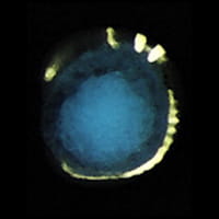
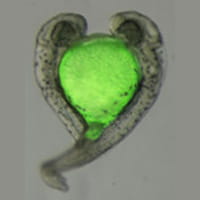
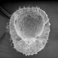
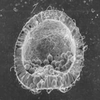
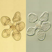
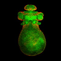
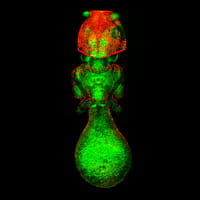
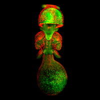
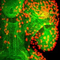
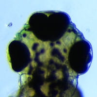
This page contains selected images that were taken during our research and training.
Overlayed image of spontaneous twin with autofluorescence in the yolk.
Anterior view of squid embryo during early development. Green is filamentous actin. Red is acetylated tubulin. Image taken during the summer of 2004 at the Woods Hole, MA, embryology course.
Anterior view of squid embryo. Green is filamentous actin. Red is acetylated tubulin. Image taken during the summer of 2004 at the Woods Hole, MA, embryology course.
Posterior view of squid embryo. Green is filamentous actin. Red is acetylated tubulin. Image taken during the summer of 2004 at the Woods Hole, MA, embryology course.
High magnification of the posterior of the squid embryo. Green is filamentous actin. Red is acetylated tubulin. Image taken during the summer of 2004 at the Woods Hole, MA, embryology course.
A spontaneously occurring partial duplication of the axis, resulting in a fish with three eyes.
Josh Waxman, PhDAssistant Professor 3333 Burnet Ave. MLC 7020Cincinnati, OH 45229
Location: S3.244Phone: 513-636-7232 Fax: 513-636-5958Joshua.Waxman@cchmc.org