The earliest that amniotic bands have been detected was at 12 weeks gestation, by vaginal ultrasound. Bands may be difficult to detect by ultrasound, and are more often diagnosed by the effect they have on the fetal anatomy, as in the case of missing or misshapen limbs.
Amniotic Band Syndrome / ABS may affect the face with cleft lip or palate, asymmetric microphthalmia or severe nasal deformity. Encephalocele may be a manifestation of Amniotic Band Syndrome / ABS, especially when eccentrically placed off the midline.
Abdominal-wall defects, typically large defects with free-floating intestine but large enough for the lines to herniate outside the abdomen, can also be the result of Amniotic Band Syndrome / ABS.
The characteristic appearance of an aberrant sheet or band of amnion attached to the fetus with resultant deformity and restriction of motion allows a diagnosis of Amniotic Band Syndrome / ABS to be made. However, prenatal diagnosis is the exception rather than the rule.
The findings in Amniotic Band Syndrome / ABS may be limited to isolated defects, including isolated facial cleft, digital amputation or mild elephantiasis of an extremity beyond a constrictive band. These features may be difficult to diagnose using ultrasound because the detailed fetal visualization required is beyond the scope of routine obstetrical ultrasound examinations.
At the worst end of the spectrum, the fetus may be so severely deformed by the amniotic bands that the spine is contracted and organs are formed in perplexing and bizarre proportions. The head may be completely misshapen or absent.
The bands responsible for these deformities are rarely seen and a presumptive diagnosis of Amniotic Band Syndrome / ABS is made based on the commonly associated deformities.
The spinal deformities in Amniotic Band Syndrome / ABS can be severe, manifesting as kyphotic lordosis or scoliosis as well as severe rotational abnormalities, even spinal amputation. While spinal deformity can be seen in other syndromes, severe spinal deformity should suggest Amniotic Band Syndrome / ABS.
Spinal deformity associated with an abdominal-wall defect is particularly suggestive of Amniotic Band Syndrome / ABS. While the typical appearance of an omphalocele is possible, the more common defect is a large slash-like defect of both the thoracic and abdominal cavities with evisceration.
These defects are associated with exteriorized bowel, liver and sometimes heart without an enveloping membrane. When associated with limb abnormalities, this is characteristic of the limb-body-wall complex form of Amniotic Band Syndrome / ABS.
Deformation of the calvarium is another group of anomalies characteristic of Amniotic Band Syndrome / ABS. If complete, the fetus may appear anencephalic. If partial, the fetus may appear to have an encephalocele.
The distinguishing features that characterize these defects as amniotic band syndrome / ABS are their asymmetric nature and associated spinal deformity or abdominal-wall defects.
In classic anencephaly, the calvarial bones are symmetrically absent. In anencephaly caused by Amniotic Band Syndrome / ABS, there is some portion of calvarium present, usually near the base of the skull or near one other orbit. Similarly, classic encephaloceles occur near the midline, while amniotic band syndrome causes encephaloceles off midline.
The presence of bands is unnecessary for the diagnosis of amniotic band syndrome / ABS in the presence of characteristic fetal anomalies. Ultrasound detection of bands is helpful in confirming the diagnosis of amniotic band syndrome / ABS as the cause of fetal deformity. However, observation of these bands without fetal abnormality is not amniotic band syndrome / ABS.
It is important for the sonographer to distinguish amniotic bands from other membranes or separations within the amnion. Separation of amnion and chorion is normal in early pregnancy until fusion occurs at approximately 16 weeks of gestation.
Chorioamniotic separation may occur as a result of amniocentesis or fetal surgery, and extrachorionic hemorrhage may separate the chorioamniotic membrane from the uterine wall. In both of these instances, a membrane may be observed by ultrasound. Other causes of membranes in the developing fetus include; septate uterus, blighted twin and circumvallate placenta.
Adhesions that form in the uterus as a result of curettage, Caesarean section, or myomectomy may cause sheets of amnion to protrude into the lumen of the amniotic cavity.
Randal et al. (1988) found that 76 percent of patients with amniotic sheets had undergone prior instrumentation. This results in an adhesion that becomes covered by chorion and amnion and has a thickness similar to the intertwin membrane of dichorionic diamniotic twins. These amniotic sheets do not adhere to the fetus because the amnion is intact.
The uterine adhesion may rupture with growth of the fetus. Filly et al. (1991) have described the ultrasound appearance of these synechiae as having a thickened base and a fine edge that undulates.
There are no associated fetal abnormalities and there is free fetal movement around the sheet. Whether due to rupture or compression by the growing fetus, the synechiae may not be seen in the third trimester.
In the limb-body-wall complex (LBWC), there is a constellation of abnormalities, including myelomeningoceles or caudal regression, thoracoabdominoschisis, or abdominoschisis and limb defects. At least two of the three abnormalities listed above are necessary to make a diagnosis of LBWC.
The umbilical cord is usually short or absent, with the placenta attached to the fetus. If present, there may be only a two-vessel cord. The limbs may be missing or the feet clubbed. The spine is often short and curved, and sacral regression is common.
There may be Arnold-Chiari malformation and hydrocephalus associated with the meningomyelocele. There may be ectopia cordis as part of the thoracoabdominoschisis. Facial clefts may also be seen.
The differential diagnosis in amniotic band syndrome / ABS depends on the ultrasound findings. In isolated constrictive amniotic bands associated with distal limb edema, possible lymphatic or vascular malformations should be considered. However, color Doppler studies should closely show the flow characteristics of a vascular malformation.
Constrictive bands involving the upper extremity should suggest the possibility of the VACTERL association, if the radius is affected, and Fanconi anemia if radial hypoplasia or absent thumbs are observed.
Amniotic membranes within the amniotic cavity, without associated fetal anomalies, may be amniotic sheets secondary to intrauterine synechiae or remnant of a blighted twin, or secondary to amniocentesis or chorionic villus sampling.
The main differential diagnosis are cases of isolated neural-tube defects or ruptured omphalocele, which do not meet the criteria for LBWC. The body-stalk anomaly has a similar constellation of anomalies, but the placenta is attached to the trunk of the fetus.
There is great controversy about the pathogenesis of the various forms of amniotic band syndrome / ABS. Part of this controversy involves the timing in gestation of the development of amniotic bands. However, in constrictive amniotic bands of the extremities, the progression of constriction combined with fetal growth has resulted in extremity amputation.
Amniotic band syndrome / ABS can be associated with either polyhydramnios or oligohydramnios. Despite the severity of some forms of amniotic band syndrome / ABS, there are no adverse maternal consequences for this diagnosis.
The incidence of intrauterine fetal death from amniotic band syndrome / ABS involving the umbilical cord is not known, but numerous cases have been reported. However, the poorly characterized pathogenesis of this syndrome and limited ultrasound surveillance limit our understanding of its natural prenatal history.
Amniotic band syndrome / ABS is a relatively common, if not always appreciated, cause of fetal and neonatal morbidity and mortality. The fetal-lamb model of Amniotic Band Syndrome / ABS has been useful in defining the pathophysiology of amniotic band syndrome / ABS and to provide a tool to understand the unique fetal response to tissue injury, repair and regeneration.
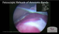
Ultrasound identification of amniotic band syndrome / ABS affecting the umbilical cord may be an indication for fetoscopic surgical intervention. Intervention for non-lethal limb deformation may also be considered for signs of threatened limb loss or evidence of umbilical cord constriction.
Constrictive bands most commonly affect the extremities, but can also involve the umbilical cord, with resulting fetal death. Kanayama et al. (1995) described the reversal of diastolic flow observed in a fetus with umbilical-cord constriction due to amniotic bands.
Graf et al. (1997) similarly reported a case of amniotic bands involving the umbilical cord following the development of chorioamniotic
separation. Despite initially normal umbilical artery Doppler waveforms, this fetus died within 2 weeks from a constrictive amniotic band of the umbilical cord.
Reports have described constrictive amniotic bands as a cause of fetal death. However, until the reports by Kanayama and Graf and their colleagues, this was a diagnosis made pathologically, after the fact. It is in cases like these that fetoscopic lysis of amniotic bands can be lifesaving.