A Novel Surgical Solution for Craniosynostosis
Published April 2021 | Neurosurgical Focus
In about one in every 2,500 births, a baby will experience craniosynostosis (premature fusion of cranial sutures). Left untreated, an abnormal head shape can increase intracranial pressure, impair intellectual development, and cause psychosocial anxiety.
Although partial fusions occur in about a third of patients with craniosynostosis, surgical literature does not include clear guidance for optimal management. Now, a novel treatment strategy is available, a technique that senior co-author Jesse Skoch, MD, describes as “taking a small surgery and making it even smaller for patients.”
Surgeons used ultrasound in the operating room to define where bone needed to be cut before opening the scalp. The surgery itself is similar to a traditional, minimally invasive, complete suturectomy, except with bone removed without endoscope and only at the point of suture fusion. Less cutting of bone, less bleeding and shorter surgery times can be achieved with this technique, all of which contributes to lower risk of longer-term consequences like failed or abnormal bone healing. This less intensive surgery was paired with postoperative helmet therapy, applied on postoperative day 4, worn for 23 hours per day for 6 to 9 months.
Five patients, average age 2.8 months, were selected for the study. Each had relatively contiguous fused regions of their cranial suture that comprised less than half the full suture. The remaining suture gaps were similar in size to the infant’s own nonpathological sutures.
Skoch says more patients need to be studied to identify the best candidates for this type of surgery. Further research will also be required to ensure the absence of relapse remains constant, but thus far, partial suturectomy with helmet therapy is suitable for very young patients and offers a less-invasive alternative to complete suturectomy with similar outcomes.
Managing Craniosynostosis
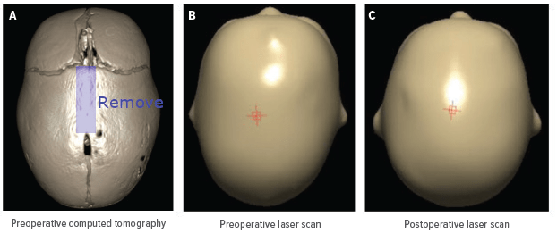
A. Preoperative CT scan of patient, demonstrating partial sagittal synostosis with phenotypical scaphocephaly. B and C: Pre- (B) and postoperative (C) laser scans of the same patient who underwent partial suturectomy for an incompletely fused sagittal synostosis.


