Diagnostic and Genetic Testing Guidance for HLH
The diagnosis of HLH is often challenging. HLH symptoms may mimic bad infections or even some kinds of cancers. Additionally, HLH can occur in patients with rheumatologic or autoimmune disorders.
Laboratory, radiologic and pathologic studies should be performed to evaluate for hidden infections or malignancies.
Laboratory evaluations can help with the diagnosis of HLH. These include:
- A cell blood count (CBC) to look for cytopenias (nearly universal)
- A routine liver panel to screen for hepatitis
- Blood levels of ferritin (usually extremely elevated), soluble IL-2 receptor (elevated), soluble CD163 (elevated), fibrinogen (decreased), and triglycerides (elevated)
- Cerebrospinal fluid analysis to indicate evidence of pleocytosis, hemophagocytosis, elevated protein and neopterin levels
Brain magnetic resonance imaging (MRI) may show evidence of HLH. Bone marrow aspirates and biopsies may show evidence of hemophagocytosis (macrophage engulfment of other cells), but this is not a required observation to make the diagnosis, nor does the absence of the sign rule out HLH.
Genetic Testing for HLH
In many cases, a genetic disorder underlies HLH.
- Some genetic forms of HLH are grouped as familial hemophagocytic lymphohistiocytosis and include mutations affecting PRF1, MUNC13-4, STXBP2, and STX11.
- Mutations in RAB27a cause Griscelli syndrome, a related disorder that may or may not be associated with pigmentary defects.
- Mutations in SH2D1A/SAP cause X-linked lymphoproliferative disease (XLP), a related disorder characterized by HLH, lymphoma and hypogammaglobulinemia.
- Mutations in XIAP cause an X-linked form of familial HLH that is often referred to as XLP2.
- Defects in LYST cause Chediak-Higashi syndrome, another related disorder characterized by HLH, pigmentary and neutrophil defects.
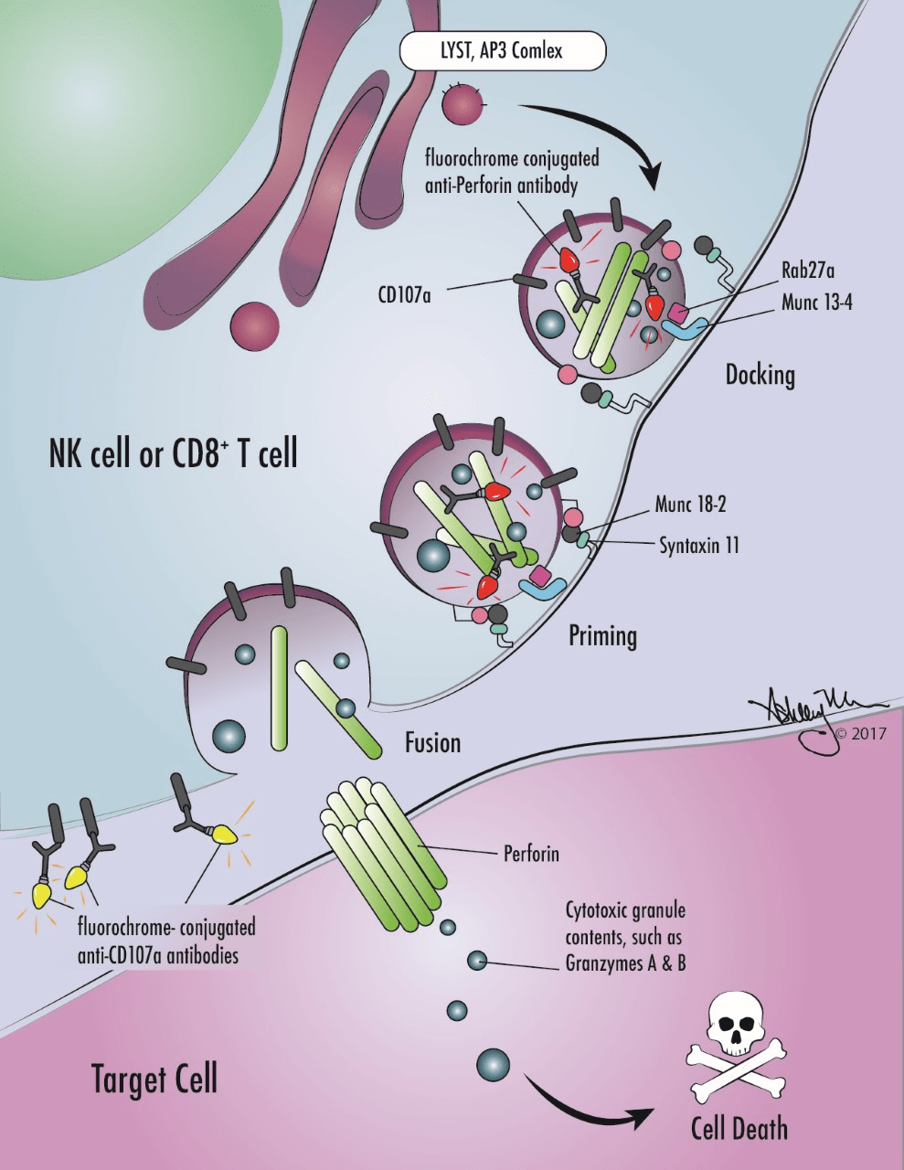
Specialized blood tests can rapidly screen patients for many of the genetic forms of HLH. Flow cytometric testing is available to screen for perforin, XIAP and SAP protein deficiencies. A functional assay to measure the degranulation of NK cells is available (termed a “CD107a assay” in our laboratory). It detects patients with defects in the genes involved in degranulation (MUNC 13-4, STXBP2, STX11, Rab27a and Lyst).
Expert Services and Guidance
If you suspect a patient has primary or secondary HLH, you can depend on Cincinnati Children’s laboratories for expert testing and clinical interpretation.
Leaders of the Diagnostic Immunology Laboratory and Molecular Genetics Laboratory supervise all aspects of the HLH diagnostic process. They offer expert advice regarding which tests to order for patients, the interpretation of results and how this data may impact treatment decisions.