What is Pectus Carinatum?
Pectus carinatum ("pigeon chest") is a common condition in which the breastbone and ribs are pushed outward. Pectus carinatum occurs more often in males than females (4:1 ratio) and develops somewhat later in males than it does in females. While it may be seen in very young children, it usually becomes more obvious during or after a growth spurt in puberty.
What causes Pectus Carinatum?
The cause of pectus carinatum is currently unknown. It tends to happen in generations of families, which suggests that genetics may play a role.
Excessive growth and structural abnormalities of cartilage (tough, connective tissue) of the ribs and breastbone are present in pectus carinatum.
What are the Signs and Symptoms of Pectus Carinatum?
For the most part, children with pectus carinatum do not have symptoms. If symptoms are present, they are typically mild, occasional chest wall pain and tenderness. But the psychosocial impact may be hard for the child. Patients with pectus carinatum often have poor body image and low self-esteem.
What are Other Disorders Associated with Pectus Carinatum?
Some children with pectus carinatum also have scoliosis (sideways curvature of the spine), kyphosis (outward curvature of the spine) or connective tissue diseases such as Ehlers-Danlos and Marfan syndromes.
Some have mitral valve prolapse, a condition where the heart mitral valve does not work normally.
How is Pectus Carinatum Diagnosed?
Some patients may have a chest X-ray during their initial evaluation. This helps the surgeon screen for other chest abnormalities or scoliosis. In severe cases of breathing problems, a pulmonary function test may help to determine how much lung function is compromised.
How is Pectus Carinatum Treated?
Fortunately, because the chest wall in children and teens is very soft and flexible, pectus carinatum usually can be corrected with bracing. Less than 10% of children with pectus carinatum need surgery to correct the problem.
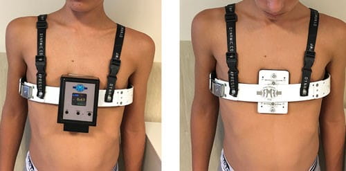
FNF Dynamic Compressor System
The FMF Dynamic Compressor System™ is a light aluminum brace that can be adjusted to fit the child. It is easily hidden under clothing. The brace gives the right amount of pressure to slowly reshape the chest wall with little discomfort.
At the first visit, a pressure device is used to measure the chest wall to decide how much pressure is needed to fix the pectus carinatum. This helps the medical team develop a treatment plan, including how many hours a day the child will need to wear the brace and for how many months.
Once a month, patients have a follow-up examination and brace adjustment. Once the pectus carinatum is corrected, patients are gradually weaned off the device to keep the shape of the newly corrected chest wall.
Advantages of the FMF Dynamic Compressor System
One of the benefits of this pectus carinatum bracing system is that it objectively measures the pressure needed to fix the condition. This measurement guides treatment decisions and helps the medical team measure progress. It also allows the medical team to give an estimate of how long treatment will last and what the chest will look like afterward. Results usually happen faster with this system compared to other bracing systems.
The success rate for the FMF Dynamic Compressor System is greater than 90% when children and teens wear it as directed. Younger children and teens, who usually have a very soft and flexible chest wall, tend to do best. In the rare event the chest wall is too stiff and does not respond to bracing, surgery to correct the pectus carinatum is still an option.


