Summary Page Tetralogy of Fallot
Normal
Tetralogy of Fallot
- Right ventricular outflow obstruction. Typically multi-level: small, malformed pulmonic valve, hypoplastic proximal pulmonary artery, and muscular narrowing below the valve.
- Right ventricular hypertrophy (thickened muscle wall) secondary to higher pressure load on this chamber.
- The aorta "overrides" the VSD.
- Ventricular septal defect. With significant obstruction in the right ventricular outflow tract, blood will shunt from right to left, bypassing the lungs and leading to cyanosis.
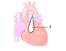
- Muscular right ventricular outflow obstruction has been cut away as part of the repair.
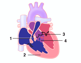
- Patch closure of the VSD.
- Right ventricular outflow patch to address all levels of obstruction.
Tetralogy repair - intracardiac view

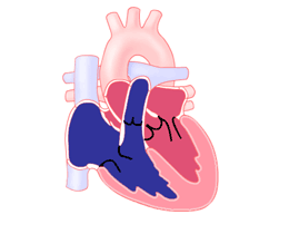
Tetralogy repair - surface view