Overcoming the Diagnostic Challenges of Infectious Diseases

Our work is dedicated to overcoming the diagnostic challenges of infectious diseases by developing innovative tools to diagnose difficult-to-detect infections, alongside the development of new treatment approaches.

Lab Projects (7)

Host-Directed Treatments for infections

Excessive and dysregulated inflammation is a well-recognized driver of poor outcomes in many infectious diseases. When the immune response becomes uncontrolled, it can cause extensive tissue damage, leading to significant morbidity and mortality. Host-directed treatments (HDTs) offer a promising strategy to address this challenge. Unlike conventional treatments that primarily target the pathogen, HDTs act on the host immune system, modulating its response to reduce harmful inflammation, limit tissue damage and also improve pathogen killing. Additionally, by targeting the host rather than the pathogen, HDTs decreases the risk of developing antibacterial resistance, an emerging global threat projected to become a leading cause of death worldwide. The overarching goal of this project is to develop and evaluate HDTs that can prevent organ damage and post-infectious complications, shorten treatment durations, and ultimately improve clinical outcomes for patients with infectious diseases, including but not limited to M. tuberculosis and S. aureus infections.

Developing Molecular imaging Approaches to Diagnose and Monitor Infections Anywhere in the Body

Deep-seated infections are challenging to diagnose and often require invasive procedures, such as biopsies, to establish a definitive diagnosis. Molecular imaging offers a promising noninvasive alternative for detecting infections and monitoring treatment response. Although 18F-FDG PET (radiolabeled glucose) is increasingly used, it lacks specificity for infections and cannot reliably distinguish infection from sterile inflammation. To address this limitation, several pathogen-specific imaging tracers targeting bacteria and fungi are being developed in Dr. Jain’s laboratory. The overarching goal is to establish a robust pipeline of pathogen-specific imaging probes that can accurately identify, localize, and monitor a broad range of infectious agents. This platform has the potential to transform the diagnosis and management of infectious diseases by enabling precise, noninvasive, and real-time assessment of infection.

Imaging to Study Antimicrobials Pharmacokinetics

Current antimicrobial dosing strategies are largely guided by achievable serum concentrations; however, growing evidence underscores the importance of measuring drug exposure directly at sites of infection. A clear example is the dosing and predicted antibiotic efficacy of anti-tuberculosis drugs used for tuberculous meningitis (TB meningitis). Despite recognition that many antibiotics penetrate the brain inadequately-primarily due to the relative impermeability of the blood-brain and the blood-cerebrospinal fluid barriers-current TB meningitis treatment regimens remain based on those developed for pulmonary TB. Dr Jain’s laboratory was the first to demonstrate the use of dynamic PET imaging to perform multi-tissue pharmacokinetic assessments of antibiotics within infected target tissues in vivo in tuberculosis. Preclinical and first-in-human PET studies using radiolabeled pretomanid (18F-pretomanid) demonstrated higher drug exposure in the brain compared to lung tissue, supporting its use in CNS TB. Similarly, discordant drug concentrations observed across different CNS compartments in preclinical models of TB meningitis highlight the limitations of relying solely on lumbar cerebrospinal fluid (CSF) measurements as a surrogate for parenchymal drug exposure in patients.

New Regimens for Implant-Associated Infections

Staphylococcus aureus is the leading cause of serious deep-seated infections, including osteomyelitis and implant-associated infections. Orthopedic implant–associated infections represent a major clinical concern, as they are particularly difficult to treat and require prolonged intravenous antibiotic treatment due to the ability of S. aureus to form biofilms, which protect bacteria from both antimicrobial agents and host immune responses. The major focus of this project is to develop new antibiotic regimens for S. aureus methicillin-resistant (MRSA) orthopedic implant–associated infections through integrated pharmacological studies and advanced animal models.

We aim to generate mechanistic insights into antibiotic pharmacology that are critical for achieving relapse-free cure. By studying clinical S. aureus strains isolated from patients with deep-seated infections, we will also assess genetic variants associated with antibiotic resistance and enhanced virulence to develop mechanistic predictors of treatment success. This data will be used to design and optimize innovative therapeutic short, oral-only antibiotic regimens for S. aureus orthopedic implant–associated infections.

Understanding Post-Infectious Complications

Excessive inflammation during infections can ultimately lead to tissue damage, organ dysfunction and post-infectious sequelae. A clear example is post-tuberculous lung disease, a condition characterized by persistent pulmonary damage following tuberculosis (TB) infection. Our overall goal is to leverage our expertise in molecular imaging, TB pathogenesis, and the clinical translation of PET ligands to develop innovative, clinically translatable imaging technologies. These tools will enable precise characterization of post-TB lung disease and its underlying biological mechanisms. We aim to identify key determinants of treatment response, including antibiotic penetration and exposure within pulmonary lesions, as well as predictors of poor outcomes that can be targeted during TB therapy to prevent long-term complications.

Integrated Platforms for Pulmonary and TB Meningitis Research

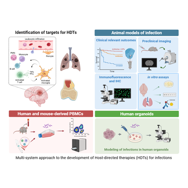
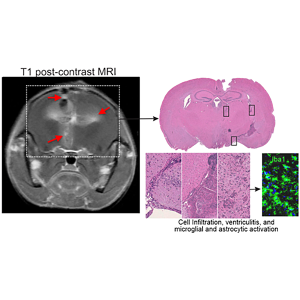
Recapitulation of the key features of human disease in animal models of tuberculosis is a critical step toward understanding pathophysiology and developing effective therapeutics. Using PET/CT, Dr. Jain demonstrated that C3HeB/FeJ (“Kramnik”) mice develop hypoxic TB lesions as well as cavitary disease—a fundamental discovery that reshaped the preclinical TB landscape and overturned the long-held dogma that matrix metalloproteinase-1 is the critical determinant of cavitation. “Kramnik” mice are now widely used in TB research worldwide, and new mechanisms underlying necrosis and cavitation continue to be explored. Similarly, a well-established model of TB meningitis has been developed in Dr. Jain’s laboratory that recapitulates key hallmarks of human disease. This model has been integrated with complementary platforms, including human brain organoids, primary human cell cultures, and advanced in vivo imaging. Using this multidisciplinary approach, we evaluate the biodistribution and penetration of TB drugs within infected brain tissues and optimize therapeutic regimens. In parallel, we investigate how intracerebral inflammatory responses and local antimicrobial exposure influence treatment efficacy, while also testing novel host-directed therapies (HDTs) that modulate neuroinflammation and reduce cerebral injury. By correlating drug distribution, host responses, and clinical outcomes, our goal is to inform the development of more effective, targeted therapies for this devastating form of tuberculosis.

Bacteria-Mediated Tumor Therapy and Molecular Imaging

Certain bacteria can naturally, or be engineered to, preferentially proliferate within hypoxic and necrotic regions of solid tumors, where conventional therapies often have limited penetration. Bacteria-mediated tumor therapy is a promising strategy to selectively target tumors, stimulate anti-tumor immune responses, and deliver therapeutic payloads such as toxins, cytokines, or enzymes. Engineered strains can also convert non-toxic prodrugs into active chemotherapeutic agents specifically within the tumor microenvironment. Overall, this approach aims to improve therapeutic efficacy while reducing systemic toxicity by minimizing delivery to non-target tissues. Additionally, bacterial colonization can be further exploited for molecular imaging to assess drug delivery and monitor treatment response. The primary goal of this exploratory project is to gain insight into the role of active targeting in increasing bacterial accumulation within tumors while using reduced systemic doses—an essential step toward improving safety for this vulnerable cancer patient population.