Health Library
Pectus Excavatum

What is Pectus Excavatum?

Pectus excavatum is a depression caused when the sternum (breastbone) is abnormally pushed inward. The depression in the chest is due to abnormal growth of the cartilage that attaches the sternum to the ribs.

Because of the deep depression, the lower ribs can stick out and give the appearance of a potbelly in younger children. This is call “rib flaring". If both sides of the breastbone are depressed in an equal fashion, the defect will look balanced. However, in many cases the chest wall appears unequal, with one side being wider.

Pectus excavatum occurs in one in 300-400 children with male prominence (male-to-female ratio of 3:1). It may be minimal, with only slight depression of the chest, or it may be quite severe. When severe, it pushes down on the heart and lungs and makes it hard for them to work properly. The abnormality often increases with age and often worsens during the growth spurts that occur during late childhood and adolescence. It usually stabilizes after skeletal growth is complete.

Causes of Pectus Excavatum

While the cause of this abnormality is unknown, the fact that it tends to occur in families suggests that genetics may play a role.

Excessive growth and structural abnormalities of the cartilage (tough, connective tissue) of the ribs and breastbone are present in pectus excavatum.

Also, pectus excavatum is seen in some inherited connective tissue disorders such as Marfan syndrome, homocystinuria, and Ehlers-Danlos syndrome.

Signs and Symptoms of Pectus Excavatum

Although some children have no symptoms, symptoms usually vary with the severity of the abnormality. Younger children have fewer symptoms than older children.

  • Children with less severe defects often have mild breathing problems.
  • Children with moderate to severe pectus deformities often have trouble breathing with exertion and are not able to tolerate exercise due to impaired lung and/or heart function. Lung capacity is decreased, and the filling capacity of the heart chambers is restricted due to compression from the sternum. These symptoms can be quite severe at times and may limit the child's activity level.
  • Growing adolescents may also experience chest pain in the area of rib cartilages.

Associated Conditions

Some children (15 percent) with pectus excavatum also have scoliosis (curvature of the spine).

Some (15 percent to 20 percent) have mitral valve prolapse, a condition in which the heart mitral valve functions abnormally.

Connective tissue disorders involving structural abnormalities of the major blood vessels and heart valves are also seen.

Although rarely seen, some children have other connective tissue disorders, including arthritis, abnormal growth, visual impairment, and healing impairment.

Diagnosis of Pectus Excavatum

Diagnostic imaging tests such as cardiac magnetic resonance imaging (MRI) is done to define the anatomy of the chest. The cardiac MRI has become choice over the CT scan. This is becomes the MRI shows cardiac involvement without requiring an echo and has no radiation exposure.

An electrocardiogram (ECG or EKG) and an echocardiogram (echo) may be performed. An ECG is a test that records the electrical activity of the heart, showing abnormal rhythms and detecting heart muscle stress. An echo is a procedure that uses sound waves to study the structures and function of the heart.

To check for breathing problems related to pectus excavatum, your child may have a breathing test done called a pulmonary function test (PFT).

Treatment of Pectus Excavatum

Sometimes mild abnormalities can be corrected with a non surgical vacuum bell treatment. Patients with moderate to severe abnormalities may require surgery, which is usually not done in children younger than age 8. Since children have softer cartilage and more flexible bones than adolescents, the operation is easier to perform on them and the cosmetic results are superior. However, good outcomes are now being obtained even in young adults.

Infants and very young children are usually not considered for surgery unless very severe abnormalities or other illnesses necessitate earlier surgery.

Surgery

Both traditional open surgery (Ravitch repair) and minimally invasive techniques (Nuss repair) are used to correct pectus excavatum. Both techniques generally improve chest appearance. They also improve the structure of the ribs and sternum, as well as the function of the heart and lung. Both techniques result in a return to normal activity and improved exercise tolerance within the first few months following surgery.

Deciding which surgical approach to use is based on a number of factors. These factors include:

  • The surgeon's level of expertise with each of these two techniques
  • The severity of the defect and its symmetry (in the middle or more to one side)
  • The child's age

Minimally Invasive Surgery (Nuss Procedure)

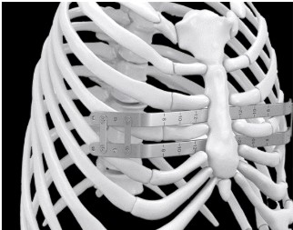
Minimally invasive surgical techniques have been used for more than three decades. With this approach, two small lateral incisions are made. A bar that has been shaped to the desired chest contour is inserted into and across the chest and positioned below the sternum. This is done using the guidance of an endoscope (instrument used to visualize the inside of the chest). The bar is held in place by sutures and by a small metal plate that prevents rotation of the bar as the chest is reshaped.

Length of hospital stay is usually two days. Children often experience more discomfort initially with the minimally invasive procedure. Postoperative erector spinae (ES) catheters analgesia catheters may be required for several days following surgery. Milder pain is managed with oral pain medication. Activity is slowly resumed, with limitations on exercise until the bar is healed in place.

With this approach it is left in place for at least three years if the bar was placed when the patient was younger than 18 years old. If placed in a patient older than 18, the bar is left in place for a minimum of five years, allowing the chest to remold. The bar is then removed during outpatient surgery.

In our experience, short-term outcomes with minimally invasive surgery are at least comparable to outcomes with open surgery. They result in a better cosmetic outcome with less scarring (due to smaller incisions), good restoration of normal chest contour, and good muscle function. Long-term outcomes currently appear to be as good as those with traditional open surgery.

What Can Be Expected Following Surgery?

There is no need to restrict exercise or activity after the initial healing period. Patients are encouraged to exercise in order to improve overall chest growth and contour and to increase the strength of their chest muscles.

Last Updated 06/2022

Patient stories.

Patients in the Chest Wall Center share their stories of hope and inspiration.

Contact us.