Newsroom
Cincinnati Children’s Images First Clinical Patient in North America Using Novel Xenon Gas
Thursday, May 11, 2023
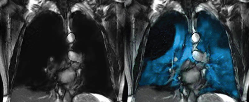
An image of a Proton MRI scan (left) and an Xe MRI scan (right).
Cincinnati Children’s Hospital Medical Center has become the first hospital in the nation to image a patient with XENOVIEWTM, a novel xenon (Xe) gas blend that enables a safe and detailed view of lung ventilation, since its FDA approval for ages 12 and older in December of 2022.
Nearly 750 children per 100,000 are affected by some type of pulmonary condition such as cystic fibrosis, asthma, or bronchiolitis obliterans. Many of these conditions are detected using a CT scan, a critical tool which, unfortunately, presents a small safety risk in the form of radiation exposure. Until now, there has not been a way to evaluate regional lung function and ventilation quickly and accurately that poses minimal potential harm to pediatric patients.
Jason Woods, PhD, director of research in the division of Pulmonary Medicine at Cincinnati Children’s, has been investigating a way to alleviate these concerns. For more than 20 years, he has researched Xe gas to pioneer a safer, faster, and more precise method for pinpointing lung disease. This research culminated in the launch of the Xenon-129 MRI Clinical Trials Consortium in 2015 which began exploring the potential of hyperpolarized Xe gas imaged via radiation-free magnetic resonance imaging (MRI) as a standardized tool at other sites.
"Xe MRI is a safer diagnostic test that helps us better understand where the problem spots are for pediatric patients,” says Woods. “It can potentially guide us to the best treatment and help us determine whether a therapy is effective. This type of imaging gives us 3D visualization, so we know how to better intervene with medications or procedures. Xe MRI is a large step forward in assessing lung ventilation.”
The Xe MRI dose is created through a Polarean HPX hyperpolarization system and administered to the patient in a small delivery bag. Once inhaled, the gas is distributed throughout the lungs and the pattern is imaged via MRI, providing a detailed look at lung ventilation and obstruction. The entire procedure takes place in a single 10- to 15-second breath hold. The gas is then exhaled and begins to leave the body within seconds.
“The gas increases regional sensitivity and specificity and provides, for the first time, a detailed map of regional ventilation as opposed to a global measure. This enables experts to assess the severity of disease and precisely where disease worsens over time, or where and how it improves with treatment,” says Woods. “In a landscape of increasingly personalized pharmaceuticals, we believe this will allow both the patient and provider to understand these changes, improving care delivery.”
Widespread use of this test could introduce a sea change in pediatric pulmonary care. In the coming years, Woods and the Consortium will continue to promote implementation to make Xe MRI a standard pulmonary diagnostic test.
3333 Burnet Avenue, Cincinnati, Ohio 45229-3026
© 2026 Cincinnati Children's Hospital Medical Center. All rights reserved.


