Wells Lab
Generating Human Tissues from Pluripotent Stem Cells

Generating Human Tissues from Pluripotent Stem Cells

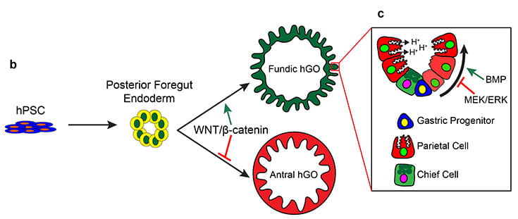
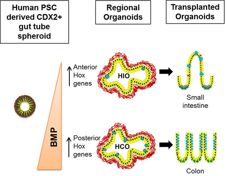
Using the signaling pathways that control organ development in vivo, we can direct the in vitro differentiation of pluripotent stem cells into human organ tissues called organoids. Organoids can be used to study human organ development or can be used to study congenital and infectious diseases. Examples include gastrointestinal (GI) tissues (esophagus, stomach, intestine, colon) that are used to study malabsorption syndromes and infectious diseases of the GI tract and endocrine cells, including pancreatic beta cells that we use to study genetic forms of diabetes. 

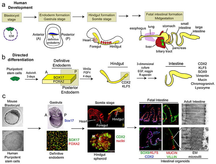
Differentiation of Human PSCs into Intestinal Tissue (Spence et al., 2011. Nature)

Studies in embryonic development have guided successful efforts to direct the differentiation of human embryonic and induced pluripotent stem cells (PSCs) into specific organ cell types in vitro. For example, human PSCs have been differentiated into monolayer cultures of liver hepatocytes and pancreatic endocrine cells that have therapeutic efficacy in animal models of liver disease and diabetes respectively. However the generation of complex three-dimensional organ tissues in vitro remains a major challenge for translational studies. We have established a robust and efficient process to direct the differentiation of human PSCs into intestinal tissue in vitro using a temporal series of growth factor manipulations to mimic embryonic intestinal development. This involved activin-induced definitive endoderm (DE) formation; FGF/Wnt induced posterior endoderm pattering, hindgut specification and morphogenesis; and a pro-intestinal culture system to promote intestinal growth, morphogenesis and cytodifferentiation. The resulting three-dimensional intestinal “organoids” consisted of a polarized, columnar epithelium that was patterned into villus-like structures and crypt-like proliferative zones that expressed intestinal stem cell markers. The epithelium contained functional enterocytes, as well as goblet, Paneth, and enteroendocrine cells. Using this culture system as a model to study human intestinal development, we identified that the combined activity of Wnt3a and FGF4 is required for hindgut specification whereas FGF4 alone is sufficient to promote hindgut morphogenesis. Our data suggests that human intestinal stem cells form de novo during development. Lastly we determined that NEUROG3, a pro-endocrine transcription factor that is mutated in enteric anendocrinosis, is both necessary and sufficient for human enteroendocrine cell development in vitro. In conclusion, PSC-derived human intestinal tissue should allow for unprecedented studies of human intestinal development and disease.

Human Intestinal Development

Figure. Model comparing embryonic intestinal development versus directed differentiation of human PSCs into intestinal tissue in vitro.

a, Schematic of human intestinal development. At the blastocyst stage, the inner cell mass (ICM) gives rise to the entire embryo. The ICM is also the source of embryonic stem cells. At the gastrula stage, the embryo contains the three germ layers including the embryonic/definitive endoderm (yellow). The definitive endoderm forms a primitive gut tube, with the hindgut forming in the posterior region of the embryo. The hindgut undergoes intestinal morphogenesis forming the small and large intestines. b, Schematic of directed differentiation of PSCs into intestinal tissue. PSCs cultured for 3 days in ActivinA form definitive endoderm (DE) co-expressing SOX17 and FOXA2. DE cultured for 4 days in FGF4 and Wnt3a (500ng/ml each) form three-dimensional hindgut spheroids expressing the posterior marker CDX2. Spheroids formed intestinal organoids when grown in three dimensional conditions that favor expansion and differentiation of intestinal precursors (matrigel with 500ng/mL R-Spondin1, 100ng/mL Noggin and 50ng/mL EGF.  c, Side-by-side comparison of embryonic intestinal development (top) and human intestinal organoid development (bottom). PSCs underwent staged differentiation in a manner that was highly reminiscent of embryonic intestinal development and formed intestinal tissue. Stages of development in c are meant to approximate the ones schematically shown in a and b

Differentiation of Human PSCs into Gastric Tissue (McCracken et al., 2014. Nature; McCracken et al., 2017. Nature)

There are two main regions of the human stomach: the anterior corpus/fundus and the posterior antrum. In the corpus, the epithelium is organized into fundic glands that contain acid-secreting parietal cells, mucous-producing cells at the surface pit and in the gland, pepsinogen-secreting chief cells, SOX2+ stem cells, endocrine cells, and rare tuft cells (8). The antrum gland unit also contains mucous cells on the surface and in the gland, endocrine cells, including gastrin-producing cells that are unique to the antrum, and rare tuft cells, but, depending on the species, may or may not contain parietal or chief cells.

To generate gastric tissues from pluripotent stem cells in vitro, it is important to utilize and temporally mimic signaling pathways that control stomach development in vivo. This involved the generation of definitive endoderm, then specification of the anterior endoderm that gives rise to the foregut through inhibition of BMP. To generate posterior foregut, requires activation of retinoic acid signaling. Additional culture with retinoic acid, followed by 3 weeks of culture in the trophic factor EGF promotes development of antral gastric organoids (McCracken et al., 2014. Nature).

The generation of fundic organoids was considerably more challenging as the signaling pathways that pattern the fundus were unknown. As described above, posterior foregut organoids and mouse genetics were used to identify that fundic specification required canonical Wnt signaling. The pro-fundic role of Wnt is separate from its earlier developmental role as a repressor of anterior endoderm, exemplifying the concept that signaling pathways have distinct roles at different stages of development. Sustained activation of Wnt signaling in posterior foregut cultures was sufficient to promote a fundic epithelial fate, resulting in the formation of fundic organoids that expressed mucous and chief cell markers. However, this was not sufficient to promote differentiation of parietal cells, which required an additional step involving inhibition of MEK and activation of BMP in the final stages of culture (McCracken et al., 2017. Nature).

Differentiation of hPSC to Gastric Tissue

Differentiation of Human PSCs into Colonic Tissue (Munera et al., 2017. Cell Stem Cell)

The generation of gastric and small intestinal organoids from pluripotent stem cells (PSCs) has greatly advanced the study human gastrointestinal (GI) development and disease. However, efforts to generate human large intestinal organoids from PSCs have lagged behind.

In effort to generate human colonic organoids, we first identified a marker, Satb2, that marks the developing large intestine throughout development and postnatally. We then identified that BMP signaling is necessary for establishing the presumptive large intestinal/Satb2+ domain of the developing gut tube of frog and mouse embryos.

Lastly, we used manipulation of BMP signaling to direct human PSC-derived gut tube cultures into colonic organoids (HCOs) in vitro. Following transplantation into mice, HCOs underwent morphogenesis and maturation forming tissue with molecular, cellular and morphologic properties of the human colon. We conclude that an evolutionarily conserved, BMP-dependent pathway patterns the human hindgut into colonic organoids, which can be used in future studies of colitis and colon cancer.

Differentiation of hPSC to Colonic Tissue