Model System

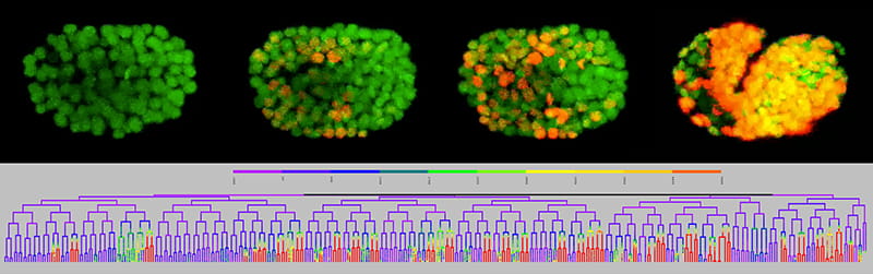
We utilize an innovative live-imaging approach that lets us measure the expression of a gene or other reporter with single cell resolution in all cells throughout development. We study the development of the tiny, but highly conserved, nematode, C. elegans, an excellent genetic model system. When combined, our approach and model system have some powerful scientific advantages. 

What is C. elegans?

Caenorhabditis elegans is a free-living (non-parasitic) worm approximately 1 mm long. They are visible to the naked eye but we use microscopes to visualize them better—both the embryo and adult are transparent. In the wild, they eat the bacteria that grow in rotting fruit and vegetation; in the lab, we grow them on agar plates with their food source, so they are very inexpensive to maintain. C. elegans are self-fertilizing hermaphrodites that complete the life cycle from fertilized egg to fertile adult in only three days, and males occur occasionally in the population which makes genetic crosses easy and fast. We can even freeze strains to save for future use.  

C. elegans was the first animal to have its complete genome sequenced in 1999. It has just over 20,000 genes, similar to the number of genes in the human genome and many of these genes are conserved by evolution, particularly those involved in development. The adult C. elegans has only 959 somatic cells, which include neurons, muscle, skin, and intestinal cells with similar functions and gene expression to those found in humans. 

What is the advantage of C. elegans in developmental biology?

For developmental biologists, the biggest advantage of C. elegans is its “invariant lineage”. This means that every embryo develops with the exact same pattern of cell divisions and cell migrations and hatches with the exact same complement of 558 cells. Larger, more complex embryos like vertebrates have millions more cells, so their development is more variable; the small size of the worm embryo both enables and requires precision. Because of the invariant lineage, we can predict the future for a given cell: we know what fate it will adopt before it starts to express any markers of its fate. 

lineage example

The invariant lineage of the C. elegans embryo was first mapped by John Sulston and published in 1983. As part of his careful work, he discovered that 113 cells undergo programmed cell death during development. The fact that some cells were “born to die” was very surprising to scientists at the time and later led to new cancer treatments that work by activating this cell death pathway to kill cancer cells. Sulston was awarded the 2002 Nobel Prize in Physiology or Medicine for his findings, along with Sydney Brenner and Bob Horvitz. Two other Nobel Prizes have been awarded for discoveries made in C. elegans: Marty Chalfie (Chemistry, 2008) won for the use of green fluorescent protein (GFP) to visualize cells and molecules in vivo and Andy Fire and Craig Mello (Physiology, 2006) won for their discovery of RNA interference. 

Building on past discoveries in C. elegans

In the Amanda Zacharias Lab, we take advantage of all three of these Nobel Prize winning discoveries to study how genes are regulated during embryonic development. We use time-lapse imaging to take 4D movies of developing embryos that carry two different fluorescent transgenes.

One is expressed in all cells and marks all nuclei in green, the other is a reporter of interest, which can be a fluorescently tagged protein or a transcriptional reporter in which a DNA fragment (i.e. enhancer or promoter) drives a red fluorescent nuclear protein. Custom image analysis software identifies the nuclei and divisions and reconstructs the invariant lineage tree, which is then color coded to show the quantitative level of expression of the reporter of interest in all cells throughout the early development of the embryo.

We can follow the embryo up to late comma stage, when it has 600 cells and begins to move on its own. Because the lineage is invariant and our approach is quantitative, we can directly compare expression patterns and levels between embryos and correlate expression with cell fate decisions. We can use RNAi to knock-down a gene of interest to measure how it affects a given reporter. Furthermore, because we know the physical position of the cells, we can track defective cell migrations in mutant embryos.