Higano Lab
Current Projects

Current Projects

Our mission is to focus on illustrating structural & functional cardio-respiratory abnormalities in perinatal patients. We do so by using novel, quantitative MRI techniques, with the objective of linking early-life MRI biomarkers of lung disease and subsequent disease trajectory in infants born preterm or with congenital abnormalities. Our goal is to improve our understanding and treatment of cardiorespiratory disease for young patients born prematurely or with congenital disorders.

Imaging of Regional Lung Ventilation in Infants

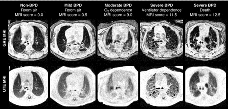
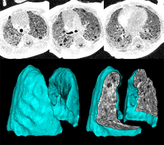
We aim to create novel imaging-based phenotyping of lung ventilation abnormalities in neonatal lung disease by employing pulmonary MRI with proton-density weighting and self-navigated gating, thus providing dynamic quantification of the 3D structure during free-breathing. Neonatal diseases of interest include bronchopulmonary dysplasia (BPD, otherwise known as chronic lung disease of prematurity) and pulmonary hypoplasia (underdeveloped lung tissue, frequently seen in congenital disorders such as congenital diaphragmatic hernia, CDH). The underlying lung abnormalities, their relationship to outcomes, and response to clinical care management is often poorly understood in patients with these conditions.

Representative axial gradient echo.

Expand image to learn more.

129Xe MRI Applications in Neonatal and Young Pediatric Lung Disease

While 129Xe inhaled gas MRI has established applications in adults and older pediatrics to investigate structure-function relationships, alveolar microstructure, and gas exchange, the technique has yet to be applied in infants. We aim to establish safety and feasibility of 129Xe MRI in infants with and without lung disease and provide clinically relevant imaging-based benchmarks for normal and abnormal pulmonary development from early infancy through the first few years of life.

Inhaled helium-3 gas diffusion MRI.

Expand image to learn more.

Phenotyping of Lung Disease of Prematurity with High Sensitivity and Minimal Risk

Obstructive and restrictive phenotypes of lung disease can be routinely measured in adults and older pediatrics using pulmonary function tests, but this procedure uncommon in infants due to technical challenges and increased patient risks. We aim to quantify surrogate metrics of obstructive and restrictive lung disease using modern quantitative 1H ultrashort echo-time (UTE) MRI in preterm neonates with bronchopulmonary dysplasia (BPD).

High-resolution 3D ultrashort echo-time MRI.

Expand image to learn more.