Walkup Lab
Current Projects

Current Projects

As highlighted by our current projects below, our ultimate goal is to improve outcomes for people living with pulmonary disease by developing new imaging-based tools to characterize lung disease pathophysiology and to individualize clinical care.

Technological and methodological developments to improve the clinical translation of Xe MRI

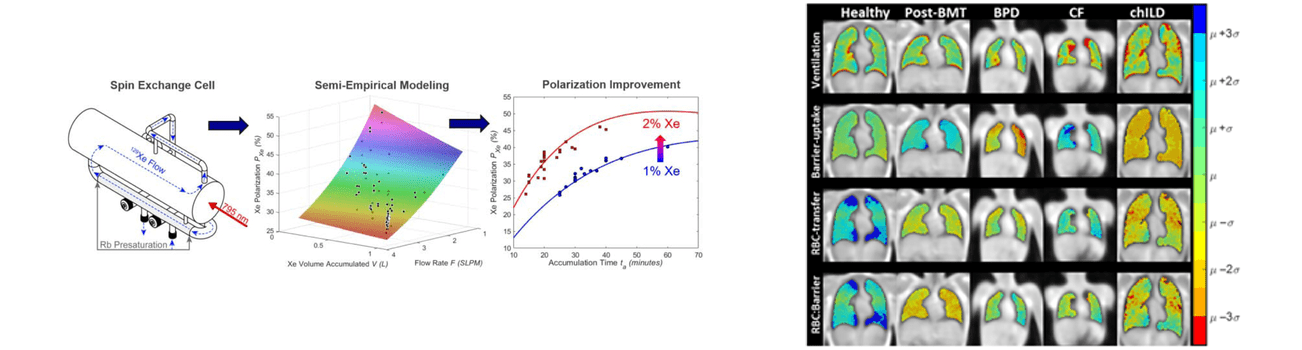
Hyperpolarized Xe gas is emerging as an inhaled contrast agent of assessing pediatric lung disease, and our group is working to improve all aspects of Xe MRI including engineering solutions for faster Xe gas production with higher MRI signal, designing MRI pulse sequences for faster acquisition with shorter breath-holds, and adapting Xe MRI techniques for younger children.

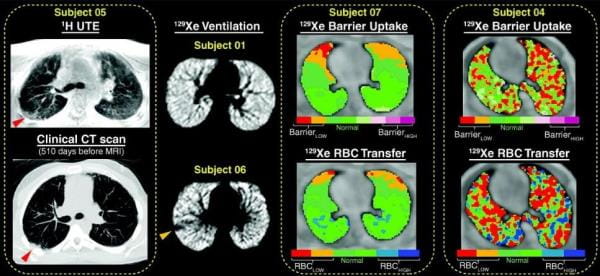
Our team was the first to demonstrate Xe MRI was safe in children and feasible even in children who are unable to perform spirometry, the clinical gold-standard test for lung disease. We recently demonstrated that Xe gas-transfer MRI, a technique that provides regional measurement of pulmonary gas exchange in adults, was feasible in pediatric populations and detected membrane-uptake and red-blood cell transfer abnormalities in children with lung disease.

Example gas-transfer map and a semi-empirical model.

Expand image to learn more.


Pulmonary complications following hematopoietic stem-cell transplantation and chemotherapy-associated lung injury in childhood-cancer survivors

Late-onset, non-infectious pulmonary complications following hematopoietic stem-cell transplant (HSCT) occur in up to half of all patients, are heterogeneous in etiology and in clinical course, and are deadly. Pulmonary disease is a significant source of morbidity and mortality in the HSCT population and also in the childhood-cancer survivor population, where treatment-related pulmonary toxicity may arise from chemotherapy and/or radiation therapy.

The objective in this project is to develop MRI techniques for early detection and phenotyping lung injury in the HSCT and childhood-cancer survivor populations. MRI outcomes may be used for risk stratification and to understand trajectories of lung injury and treatment response especially in younger children who are difficult to characterize using clinical pulmonary-function tests.

Representative axial xenon-129 (129Xe) ventilation magnetic resonance images from four haematopoietic stem cell transplantation (HSCT) patients.

Expand image to learn more.

Pulmonary structure-function relationships and regional pathophysiology in rare-lung diseases

Lung dysfunction arises from some combination of abnormal respiratory structure and function and typically, these abnormalities are diffuse especially in early disease. MRI leverages spatial resolution and is non-invasive and non-ionizing, thus there is opportunity to use MRI to understand basic pathophysiological questions about rare-diseases with a limited number of participants. For instance, we used Xe MRI to study lymphangioleiomyomatosis (LAM) and found ventilation deficits and alveolar-airspace enlargement even in participants with LAM and normal pulmonary-function tests, suggesting very early structural changes. In a case report of a child with generalized lymphatic anomaly, we used Xe MRI to determine if recurrent chylous effusions impacted alveolar growth and found normal airspace size—a question that would have required an invasive biopsy to answer. Ongoing work includes using MRI to assess treatment response in “n=1” therapeutic trials for individuals with rare- and ultra-rare lung diseases and using MRI to target biopsies to increase diagnostic yield.

Summary of notable 1H and xenon 129 (129Xe) magnetic resonance imaging findings.

Expand image to learn more.

129Xe ventilation and apparent diffusion coefficient (ADC) map.

Expand image to learn more.

Example image slices for 129Xe (a.) and UTE (b.) images in CF subjects.

Expand image to learn more.