CDH Fetal Surgery

Congenital diaphragmatic hernia / CDH is most often a posterolateral defect in the left hemidiaphragm (88%) on one side, which leads to herniation of the viscera into the thorax, resulting in pulmonary hypoplasia and respiratory embarrassment.

Congenital diaphragmatic hernia / CDH occurs in approximately 1 in 2,500 to 5,000 live births and as frequently as 1 in 2200 prenatal ultrasound studies.

This discrepancy between neonates who survive birth and transport to a tertiary newborn treatment center and fetuses diagnosed by prenatal ultrasonography supports the notion of a "hidden" mortality.

With inclusion of cases that never reach the treatment stage of disease, the mortality approaches 75%.

Although familial cases with an autosomal-dominant inheritance have been reported, most cases of congenital diaphragmatic hernia / CDH are sporadic.

Associated anomalies are seen in 25% to 57% of all cases of congenital diaphragmatic hernia / CDH and 95% of stillborns with congenital diaphragmatic hernia / CDH and include:

  • congenital heart defects
  • hydronephrosis
  • renal agenesis
  • intestinal atresia
  • extralobar sequestrations; and neurologic defects, including hydrocephalus, anencephaly and spina bifida.

Chromosomal anomalies, including trisomy 21, 18, and 13, occur in association with congenital diaphragmatic hernia / CDH in 10% to 20% of cases diagnosed prenatally.

The diagnosis of congenital diaphragmatic hernia / CDH is often an unexpected finding on routine prenatal ultrasound examination or a scan prompted by polyhydramnios.

Critical ultrasound findings include the presence of viscera in the right or the left hemithorax above the level of the inferior margin of the scapula or at the level of the four-chamber view of the heart.

The hypoechoic signal of the fluid-filled stomach, gallbladder or bowel can be distinguished from the hyperechoic signal of the fetal lung. Other common findings include:

  • A small ipsilateral lung
  • A defect in the ipsilateral diaphragm
  • A shift of the mediastinum away from the affected side

In the case of a right-sided congenital diaphragmatic hernia / CDH, the liver may be the only herniated organ, and it is difficult to distinguish it from the fetal lung because of their similar echodensities. Identification of the diaphragm does not exclude the possibility of congenital diaphragmatic hernia / CDH because some portion of the diaphragm is usually present in congenital diaphragmatic hernia / CDH.

The differential diagnosis of congenital cystic adenomatoid malformation / CCAM includes:

  • type I congenital cystic adenomatoid malformation / CCAM; bronchogenic cysts;
  • neurenteric cysts; and cystic mediastinal teratoma, which may mimic the appearance of a herniated bowel.

Identification of abnormal upper abdominal anatomy and presence of peristalsis in herniated bowel loops helps distinguish congenital diaphragmatic hernia / CDH from other diagnoses.

The location of the gallbladder in fetuses with congenital diaphragmatic hernia / CDH is helpful because it may be displaced to the midline or in the left upper quadrant or herniated into the right chest.

In most severe cases, the liver and the stomach are present in the thorax. Bowing of the portal vein or sinus venosus to the left of the midline or coursing of the portal branches to the lateral segment of the left lobe of the liver above the diaphragm can be seen with color flow Doppler imaging and is the best sonographic predictor of liver herniation.

In addition, the position of the stomach (easily seen in contrast to the more echogenic fetal lung) in a posterior or midthoracic location is also associated with liver herniation.

Several sonographic features have been suggested as prognostic indications in congenital diaphragmatic hernia / CDH, including:

  • Polyhydramnios
  • Early gestation diagnosis (less than 24 weeks)
  • Stomach herniation
  • Herniation of the left lobe of the liver
  • Evidence of fetal cardiac ventricular disproportion before 24 weeks of gestation and lung-to-head circumference ratios

However, no single sonographic feature of congenital diaphragmatic hernia / CDH has been uniformly helpful in predicting outcome. A more direct estimate of pulmonary hypoplasia which correlates with neonatal outcome is needed.

Lung area–to–head circumference ratio (LHR) (two-dimensional area of right lung measured at the level of the four-chamber view of the heart) was assessed prospectively to determine its value in predicting the postnatal outcome with conventional therapy.

The LHR, although still useful, has proven less reliable in predicting survival than was previously thought, especially in the most severe category. LHRs greater than 1.4 are still associated with an excellent survival rate in the 80 to 85% range with only 25% of patients requiring ECMO.

An LHR between 1.0 and 1.4 is associated with survival of about 75% with 69% requiring ECMO. The largest change has been the survival observed with patients with LHR less than 0.9 in which survival of up to 62% of patients can be expected but almost all survivors requiring ECMO.

It is not known if the improved survival with LHR less than 1.0 reflects improved neonatal care, such as the use of "gentilation" strategies, or if greater experience with larger numbers of patients in this category of LHR is now giving a more accurate reflection of survival.

The position of the fetal liver in left sided congenital diaphragmatic hernia / CDH remains an important prognostic factor. In our most recent experience at CHOP, survival in left sided congenital diaphragmatic hernia / CDH with liver in the abdomen was 91% with only 24% requiring ECMO.

Conversely, survival in left sided congenital diaphragmatic hernia / CDH with significant herniation of the left lobe of the liver was only 51%, with 79% of patients requiring ECMO. These statistics apply only to cases of left sided congenital diaphragmatic hernia / CDH. The LHR does not apply to right-sided congenital diaphragmatic hernia / CDH in which the liver is almost always herniated and it is not necessarily associated with a worse prognosis.

In fact in a review of the Children's Hospital of Philadelphia (CHOP) experience with 22 cases of prenatally and 6 cases of postnatally diagnosed right sided congenital diaphragmatic hernia / CDH, Hedrick et al, found an overall survival rate of 70%.

Among those diagnosed prenatally, 4 cases chose to terminate the pregnancy and of the remaining 18, 16 survived (89%, 73% if terminations are included). Half of the patients (12 of 23, 53%) required ECMO, and of those, the survival rate was 75%.

Unfortunately, none of the prognostic features which assist us in counseling patients with a left sided congenital diaphragmatic hernia / CDH apply to right-sided congenital diaphragmatic hernia / CDH.

It is noteworthy, however, that obstetrical complications including polyhydramnios, preterm labor, and premature rupture of membranes occurred in 50% of the right sided congenital diaphragmatic hernia / CDH pregnancies.

Despite the advances in neonatal care, such as high-frequency oscillatory ventilation, inhaled nitric oxide, and ECMO, the mortality rate of isolated congenital diaphragmatic hernia / CDH remains substantial.

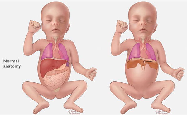
Out of frustration with these grim statistics, Harrison and colleagues pioneered fetal surgery for congenital diaphragmatic hernia / CDH. Unfortunately, survival following complete in utero repair was poor. These failures were due to herniation of the left lobe of the liver.

Reduction of the liver during repair inevitably resulted in kinking of the umbilical vein, leading to fetal bradycardia and cardiac arrest. Herniation of the left lobe of the liver became an exclusion criterion for complete in utero repair of congenital diaphragmatic hernia / CDH.

However, even if cases with left lobe herniation are excluded, the survival rate in the series by Harrison and colleagues was only 41%, which was no better than with conventional postnatal therapy at the time.

A prospective trial sponsored by the National Institutes of Health confirmed these findings; thus, there is currently no indication for complete repair of congenital diaphragmatic hernia / CDH in utero. The shortcomings of in utero repair led to development of a new approach.

Known for decades, fetal tracheal occlusion results in accelerated fetal lung growth in animal models. It was not until 1994, however, that tracheal occlusion was applied to the problem of congenital diaphragmatic hernia / CDH.

In the animal models of congenital diaphragmatic hernia / CDH, tracheal occlusion induces lung growth, increases alveolar surface area and alveolar number, as well as visceral reduction from the chest.

The results of these experiments were so compelling that fetal tracheal occlusion was applied in human fetuses with severe congenital diaphragmatic hernia / CDH.

The results of open fetal surgery for tracheal clip procedure in high-risk congenital diaphragmatic hernia / CDH were disappointing in both the UCSF and CHOP experience.

As a result of the poor outcomes with the procedure, a procedure was described using transuterine endoscopy or FETENDO. The results from the FETENDO approach in high risk congenital diaphragmatic hernia / CDH were promising and the NIH sponsored a trial of fetoscopic tracheal clip application compared to conventional postnatal therapy.

Shortly after initiation of the FETENDO trial for congenital diaphragmatic hernia / CDH the UCSF group developed a less invasive endolumenal balloon tracheal occlusion technique requiring only a single port as opposed to the 5 for the FETENDO approach. This approach, using a detachable balloon, was incorporated into the NIH sponsored trial.

The trial was halted after enrollment and randomization of 24 patients because of an unexpectedly high survival rate with standard postnatal care. The Data and Safety Monitoring Board concluded that further recruitment would not result in significant differences between the groups.

Eight of 11 (73%) in the tracheal occlusion group and ten of 13 (77%) in the group that received standard postnatal care survived. There are several important lessons to be learned from this trial.

First, these results primarily apply to fetuses with LHR > 0.9 and< 1.4.="" it="" remains="" unknown="" if="" the="" most="" severely="" affected="" fetuses="" with="" lhr="">< 0.9="" and="" liver="" herniation="" would="" do="" better="" with="" fetal="" tracheal="">

Second, the outstanding survival achieved with "standard" therapy were obtained at a tertiary center that cares for a large volume of diaphragmatic hernia patients. These results may not be generalizable to centers that do not have extensive experience caring for critically ill newborns with severe pulmonary hypoplasia due to congenital diaphragmatic hernia / CDH.

In the most severe cases of diaphragmatic hernia fetal intervention is still being investigated. In Belgium, Jan Deprest is offering fetoscopic balloon tracheal occlusion for congenital diaphragmatic hernia / CDH with LHRs less than 0.9. and herniation of the liver.

EXIT-to-ECMO

The Cincinnati Children's Fetal Care Center offers the EXIT-to-ECMO strategy as treatment for high-risk CDH.

The use of EXIT-to-ECMO was first performed by Timothy Crombleholme, MD, for the management of severe CDH (liver herniation and LHR < 0.9)="" associated="" with="" congenital="" heart="">

This has now been applied to cases of isolated high-risk CDH:

  • Left-sided CDH with herniation of the left lobe of the liver and LHR ≤ 0.9, PPLV ≤ 15, TLV ≤ 18 and karyotype with no cardiac defects
  • Right-sided CDH with PPLV ≤ 15 and TLV ≤ 18 

The rationale for this approach is that by transitioning directly from placental support to ECMO support the infant is never hypoxic, acidotic or hypotensive and is never exposed to barotrauma from vigorous neonatal resuscitation. From a maternal risk standpoint, this approach obligates the mother to a surgical delivery.

From the fetal standpoint, because this approach is reserved for high-risk CDH, no additional fetal risk is incurred. EXIT-to-ECMO is thought to improve survival in CDH with overall survivals of 65 percent in high-risk groups of patients in which survival of 11 to 40 percent is routinely expected.

For More Information

For more information or to request an appointment, contact the Cincinnati Children's Fetal Care Center at 1-888-338-2559 or use our online form.

Contact us.

Congenital diaphragmatic hernia occurs when the muscle separating the abdomen and chest cavities does not form or forms with a hole in it. Some of the organs in the abdomen push through the hole into the chest, crowding the lungs and causing them to develop poorly.
Click for larger image.