From Patterns to Organs
 |
|
Figure. This image from Spence et al. shows a section through the developing pancreas and biliary system of an E10 mouse embryo. The pancreas (Pdx1 – green) and biliary tree Sox17 (blue) derive from a common pool of ventral foregut progenitor cells. Segregation of these organ lineages depends on Sox17 and notch signaling. This C&Bs image was acquired using a Zeiss confocal microscope. |
We have several projects to investigate the mechanisms of development of specific organs including lungs, esophagus, stomach, pancreas, and intestines.
Pancreas and Biliary Development (from Spence et al., Dev Cell. 2009)
The ventral pancreas, biliary system and liver arise from the posterior ventral foregut, but the cell-intrinsic pathway by which these organ lineages are separated is not known. Here we show that the extrahepatobiliary system shares a common origin with the ventral pancreas and not the liver, as previously thought. These pancreatobiliary progenitor cells coexpress the transcription factors PDX1 and SOX17 at e8.5 and their segregation into a PDX1+ ventral pancreas and a SOX17+ biliary primordium is Sox17-dependant. Deletion of Sox17 at e8.5 results in the loss of biliary structures and ectopic pancreatic tissue in the liver bud and common duct, while Sox17 overexpression suppresses pancreas development and promotes ectopic biliary-like tissue throughout the PDX1+ domain. Restricting SOX17+ biliary progenitor cells to the ventral region of the gut requires the notch effector Hes1. Our results highlight the role of Sox17 and Hes1 in patterning and morphogenetic segregation of ventral foregut lineages.
 Cover Image of Developmental Cell
This image from Spence et al. shows a section through the duodenum and caudal stomach of an E16.5 mouse embryo in which Sox17 was ectopically expressed in the Pdx1 domain of the gut during early embryogenesis (E9-10.5). Sox17 is required for normal development of the extrahepatic biliary ducts and its misexpression leads to the development of ectopic ductal tissue (HNF6 – green) and loss of duodenal (HNF4alpha – red) and pancreatic tissue. Nuclei are counterstained with Topro3 (blue). The image was acquired using a Zeiss confocal microscope.
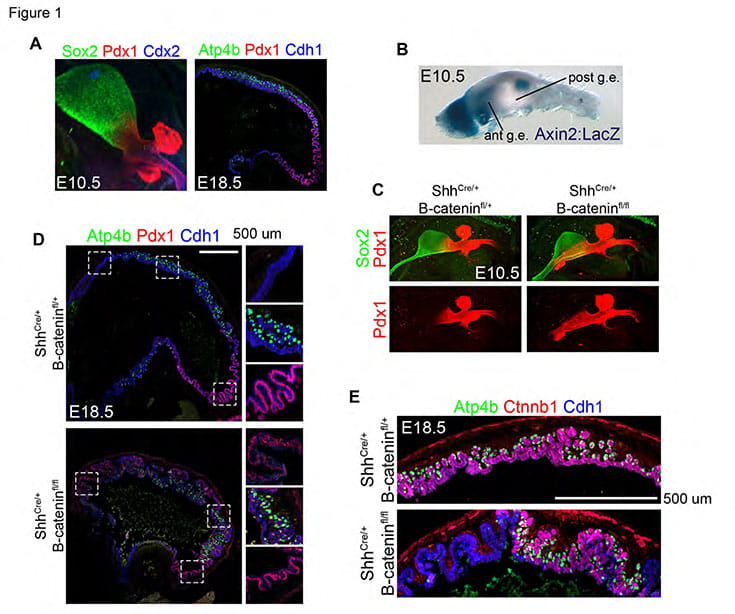
Stomach Development (from McCracken et al., 2017. Nature):
The stomach is one of the most structurally diverse organs among mammals4. In humans, the gastric mucosa generally consists of two types of epithelial glands5,6. Located in the more proximal anatomic domains – the corpus and fundus – of the stomach, oxyntic glands comprise acid-secreting parietal cells, protease-producing chief cells, mucus-producing cells, and endocrine cells. Antral-type glands, located in the more distal antrum and pylorus, contain mostly mucous and endocrine cells. The fundus and antrum derive from a common population of posterior foregut progenitors, however the mechanisms that separate these stomach domains in vivo were not previously known. We found that Wnt/β-catenin signaling is required for fundus specification in mice and that loss of the canonical Wnt signaling effector, beta-catenin, from the gastric epithelium results in antralization of the stomach. Further, disruption of this early patterning coincides with subsequent cell autonomous loss of parietal cells, suggesting that cytodifferentiation is impaired secondary to developmental patterning defects.