Current Projects
Current Projects
Project 1: The role of HLA-G+/C+ extravillous trophoblasts (EVT) in tolerance and immunity
We developed new techniques to isolate and culture HLA-G+ HLA-C+ extravillous trophoblasts from 1st trimester and term pregnancy samples. This has led to their extensive characterization as well as investigation into their interaction with decidual immune cells (decidual NK cells, decidual macrophages and CD4+ and CD8+ decidual T cells).
These studies demonstrated that HLA-G+ trophoblast can enhance the proportion of regulatory T cells (Treg) that can suppress anti-fetal responses. Further studies demonstrated that cytokine-activated decidual CD8+ effector T cells and natural killer (dNK) cells are not equipped to kill allogeneic trophoblast cells even when they are infected with bacteria or viruses. This suggests that HLA-G+/C+ trophoblast are well-equipped to deter a detrimental maternal immune response and contribute to maternal fetal tolerance.
We recently setup methods to establish highly proliferative HLA-G expressing EVT-like cell lines from human pregnancy placental samples. This breakthrough technology allows us to investigate HLA-G+ EVT differentiation in more detail and determine further molecular mechanism of EVT and lymphocyte interactions and establishment of maternal-fetal tolerance.
Collaborators:
- Dr. Emily DeFranco
- Dr. Wendy Goodman
Funding:
- 07/01/2021 – 03/31/2025 Burroughs Wellcome Fund - Next Gen Pregnancy Initiative – Award. PI: Tilburgs
- 01/01/2021 – 12/01/2023March of Dimes – Ohio Preterm birth Collaborative Role; PI: Way

Project 1: The role of HLA-G+/C+ extravillous trophoblasts (EVT) in tolerance and immunity. Confocal images of HLA-G+ extravillous trophoblasts (EVT) interacting with decidual NK cells (dNK). EVT and dNK interact through filo podia-like structures (middle panel) as well as immune synapses in which perforin does not localize to the synapse region (right panel). DAPI (blue), HLA-G (red), F-actin (green), perforin (white). Tilburgs, Evans et al., PNAS 2015 PMID: 26460007.
Project 2: Regulation of fetus- and virus-specific CD8+ T cells in human pregnancy
CD8+ T cells are the main effector cells at the maternal-fetal interface that can elicit an allogeneic response to invading fetal trophoblast cells that express polymorphic HLA-C molecules. Furthermore CD8+ effector T cells are also the most important effector cells that maintain immunity to viral infections at this site.
Dr. Tilburgs work demonstrated that human decidual tissue contains highly differentiated CD8+ effector-memory T cells that lack a clear cytolytic capacity.
Current work demonstrates that upon activation these CD8+ EM T cells become fully functional and secrete pro-inflammatory cytokines and upregulate cytolytic molecules, but they are not able to kill healthy but allogeneic trophoblasts. Current work focuses on the ability of decidual CD8+ T cells to maintain fetal tolerance while providing immunity to viral infections, in particular HCMV infection.
Funding:
07/01/2020 – 06/30/2022 Cincinnati Children's Research Foundation - Trustee Award. PI: Tilburgs
Collaborators:
- Dr. Sandra Andorf
- Dr. Krishna Roskin
- Dr. Clive Gray and Dr. Nadia Ikumi, University of Cape Town, Cape Town, South Africa

Project 2: Regulation of fetus- and virus-specific CD8+ T cells in human pregnancy. Decidual CD8+ T cells are differentiated effector-memory cells but unlike blood CD8+ EM cells decidual CD8= EM T cells express very low levels of the cytolytic molecule perforin (Tilburgs et al., J. Immunol 2010). Further gene expression analysis demonstrated that of decidual CD8+ T cells have increased signature of activation and dysfunction possibly to accommodate the conflicting demands establishing tolerance and providing immunity to infection (van der Zwan et al., PNAS 2018; Papuchova et al. Front. Immunol. 2019).
Project 3: Decidual NK cell response to infection
For the longest time, decidual NK cells were considered low cytotoxic cells with unique capacities to secrete cytokine and growth factors to promote trophoblast invasion and placental development. Despite dNK high expression of cytolytic granules (perforin, granzyme B, and granulysin), their cytolytic responses to pathogen infected cells was not studied.
Dr. Tilburgs and colleagues demonstrated that dNK are able to kill virus (HCMV) infected decidual stromal cells but not HCMV-infected fetal trophoblasts, suggesting that viral infections are not well-controlled in the placenta.
Moreover, expression of the activating NK receptor KIR2DS1 on dNK and its interaction with HLA-C increased dNK degranulation and leads to enhanced immunity. This may explain how KIR2DS1 reduces risk for pregnancy complications as was shown by Hiby and Moffett et al., in genetic studies.
Other projects include the novel finding that dNK selectively transfer granulysin to kill bacteria inside trophoblasts without killing the trophoblasts and the unique differences found between decidual NK cells in early pregnancy and at term pregnancy. Key research questions are to determine whether separate populations of dNK responsible for placental development and immunity exist and how dysfunction of dNK contributes to pregnancy complications including maternal to fetal transmission of pathogens.
Collaborators:
Dr. Christian Karsten, University of Lübeck, Germany
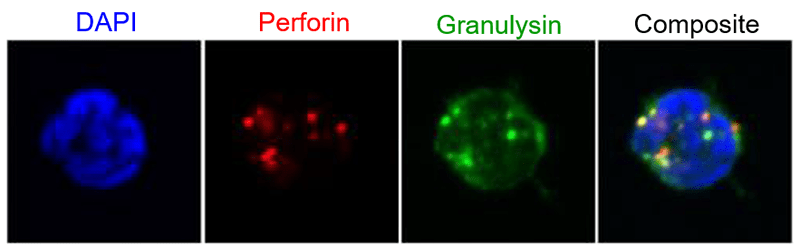
Project 3: Decidual NK cell response to infection. Confocal image of decidual NK cell filled with the cytolytic granules perforin (red) and granulysin (green).
Project 4: The role of regulatory T cells at the maternal-fetal interface in human pregnancy
During her Ph.D. studies at the Leiden University Medical Center in Leiden, Netherlands (2004-2008), Dr. Tilburgs was the first to discover that fetus-specific HLA-C recognition by maternal T cells occurs at the human maternal-fetal interface in healthy pregnancies. However, the increased T cell activation is accompanied by the induction of functional T regulatory cells that can suppress this response.
In further research, the Tilburgs Lab demonstrated that HLA-G+/C Extravillous trophoblasts (EVT) directly increased the percentage of FOXP3+ Treg as well as the FOXP3 protein levels in these cells. Recently, publications have identified additional decidual Treg subtypes with functional suppression capacity and described what type of immune responses these Treg types can suppress and how HLA-G+ EVT and decidual macrophages induce these Treg.
The current project focus is on the function and dysfunction of Treg populations during pregnancy complications, as well as their specificity to fetal antigens.
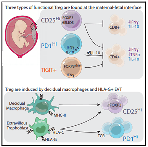
Project 4: The role of regulatory T cells at the maternal-fetal interface in human pregnancy. Salvany-Celades et al. Cell Reports 2019.
3333 Burnet Avenue, Cincinnati, Ohio 45229-3026
© 2026 Cincinnati Children's Hospital Medical Center. All rights reserved.


