Parikh Research Lab
The goal of the Parikh Lab is to enable early detection, at birth, and prevention/treatment of neurodevelopmental disabilities.
Increased survival of extremely preterm infants has contributed to a higher prevalence of survivors with motor, cognitive, and behavioral/psychiatric abnormalities. Accurate diagnosis of these abnormalities takes 2 to 3 years. These early years are when the brain is most active in building its wiring system and optimally receptive to change and healing.
Thus, when the diagnosis is delayed by up to 3 years, precious time is lost. Newer approaches to diagnosis, prediction and prevention of developmental disabilities are urgently needed to improve the long-term quality of life of high-risk newborns.
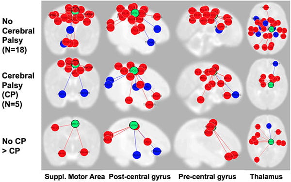
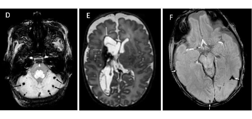
The Parikh Lab employs advanced brain MRI tools such as volumetric, diffusion, and functional MRI for early identification of biomarkers of brain injury/delayed development that are predictive of disabilities in individual high-risk neonates/infants. For example, structural and functional connectivity biomarkers are poised to meet this critical need.
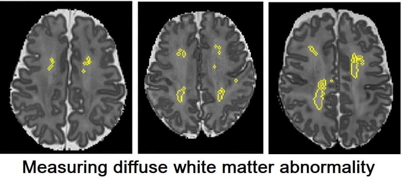
The lab’s current focus is to understand the nature of the commonly encountered diffuse white matter abnormalities and to develop early prognostic models of motor, cognitive, and behavioral abnormalities in a geographic cohort of 350 very preterm infants – The Early Prediction Study. In addition to using multivariable regression modeling, the study is employing newer machine learning/artificial intelligence approaches to uncover novel prognostic biomarkers to enhance outcome prediction of neurodevelopmental disabilities. This important step will facilitate risk stratification/early detection, at birth, to design clinical trials of targeted neuroprotective interventions during the critical window of the first 3 years after birth when brain plasticity is at its peak.


