Ventral Body Wall Formation
Midline defects of the body wall account for approximately 5% of congenital abnormalities observed at birth. Defects in the closure thoracic body wall result in conditions such as ectopia cordis (exposed heart) and anomalous sternum affecting the normal development of the lungs. The molecular mechanisms underlying the formation of the ventral body are poorly understood.
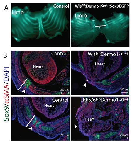
We are currently studying models wherein perturbation to Wnt ligand secretion, or reception impairs the thoracic body wall's closure, resulting in a malformed sternum, exposed heart, and abnormal pulmonary morphology. These studies will provide insights into normal body wall development and abnormal body wall closure pathogenesis.