Patient and Family Resources
Comprehensive, Compassionate, and Respectful Care for Patients and Families
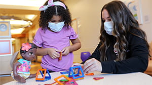
Our team of experts offers individualized care and attention to match your child’s unique needs, and we provide a variety of resources to ensure your child and family receive the support you need during your time with us.
To make your visit and treatment a smooth process, we offer a number of online resources. Use the following links to learn more about our services and resources available to help your child and family:
Prepare for Your Visit


Additional Resources
3333 Burnet Avenue, Cincinnati, Ohio 45229-3026
© 1999-2025 Cincinnati Children's Hospital Medical Center. All rights reserved.


