Pulmonary Biology
Lung Morphogenesis

Lung Morphogenesis

Lung dysfunction at birth can occur due to prematurity or a number of congenital problems. Since perinatal death is most frequently associated with lung dysfunction, many of the groups in our division study the molecular and cellular mechanisms and processes that regulate lung development (morphogenesis). Lung morphogenesis occurs both prenatally and postnatally and is typically divided into five phases (see figure 1), with the final alveolar phase occurring principally after birth in humans and rodents. Our groups work on the pathways regulating each specific developmental phase, as well as the different cellular and structural processes distinct to each phase. As well as these important development pathways, lung maturation is crucial as it prepares the lung for birth, when the lung must very rapidly function as the gas exchange organ for the body. Prior to birth the fetus receives oxygen via the mother’s placenta. Surfactant is critical for lung function at birth, reducing air-liquid tension and allowing for lung expansion. Our division has a long history in understanding the role of different surfactant components and surfactant biology and regulation. For more information on these and the other developmental processes listed below, visit the faculty lab websites and biosketches below.

Lung morphogenesis studies in the Division of Pulmonary Biology include:
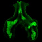
 

Early Lung Specification and Morphogenesis.

Detection of E-cadherin by immunofluorescent staining shows epithelial cells & branching in early lung morphogenesis. (Picture courtesy of the Shannon laboratory.)


Vrushank Davé, PhD: Transcriptional/signal networks in lung development and cancer 

Alan Kenny, MD, PhD: Molecular mechanisms early respiratory and gut development

Anne-Karina Perl, PhD: Lung repair processes and progenitor cells in lung development

John Shannon, PhD: Lung morphogenesis and differentiation, and FGFs

Susan Wert, PhD: Histopathology of abnormal lung development, injury / repair and human surfactant dysfunctions / mutations.

Jeffrey Whitsett, MD: Lung transcriptional control, epithelial patterning and differentiation

Yan Xu, PhD: Bioinformatics, systems biology of lung development and disease

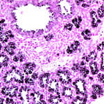
 Epithelial Cell Patterning and Differentiation. Immunostaining for pro-surfactant protein C (black) identifies type II epithelial cells in the distal saccules of developing lung of a transgenic mouse at E17.0.


Vrushank Davé, PhD: Transcriptional/signal networks in lung development and cancer

Machiko Ikegami, MD, PhD: Lung development and C/EBPα

Anne-Karina Perl, PhD: Lung repair processes and progenitor cells in lung development

John Shannon, PhD: Lung morphogenesis and differentiation, and FGFs

Susan Wert, PhD:   Histopathology of abnormal lung development, injury / repair  and human surfactant dysfunctions / mutations.

Jeffrey Whitsett, MD: Lung transcriptional control, epithelial patterning and differentiation

Yan Xu, PhD:  Bioinformatics, systems biology of lung development and disease


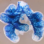
 Epithelial-to-Mesenchymal Signaling Mechanisms. Vascular morphogenesis in the developing lung is regulated by a complex system of epithelial-to-mesenchymal signaling mechanisms. Picture shows X-gal staining of lungs form Tie2-Lac Z mice at E12, which detects the endothelial plexus, a primitive vascular network that is forming alongside the developing airways. (Picture courtesy of Dr. Bridges in the Shannon laboratory.)


James Greenberg, MD: Lung vascular, lymphatic, airway development and role of VEGF

Timothy Le Cras, PhD: Lung development, pathogenesis of lung remodeling

John Shannon, PhD: Lung morphogenesis and differentiation, and FGFs

Jeffrey Whitsett, MD: Lung transcriptional control, epithelial patterning and differentiation


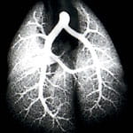
 Vascular Development. Arteriogram shows complex system of branches that develops in the lung to supply blood for oxygenation to the distal airspaces (alveoli). (Picture courtesy of the Le Cras laboratory.)


James Greenberg, MD: Lung vascular, lymphatic, airway development and role of VEGF

Timothy Le Cras, PhD:  Lung development, pathogenesis of lung remodeling

Jeffrey Whitsett, MD:  Lung transcriptional control, epithelial patterning and differentiation


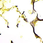
 Alveolar Morphogenesis. Picture shows elastin fibers (black) in the walls of alveoli and at the tips of secondary septae in the lung. The correct deposition of elastin fibers is critical for alveologenesis and alveolar structure. Alveoli are the functional site of gas exchange in the lung and form the large surface area of the lung needed for gas exchange. (Picture courtesy of the Le Cras laboratory.)


Timothy Le Cras, PhD: Lung development, pathogenesis of lung remodeling

Anne-Karina Perl, PhD: Lung repair processes and progenitor cells in lung development

John Shannon, PhD:  Lung morphogenesis and differentiation, and FGFs

Susan Wert, PhD: Histopathology of abnormal lung development, injury / repair  and human surfactant dysfunctions / mutations.

Jeffrey Whitsett, MD: Lung transcriptional control, epithelial patterning and differentiation


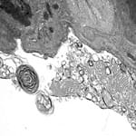
 Surfactant Regulation and Biology. Electron micrograph shows that surfactant in the airspace is present in multiple forms including lamellar body, tubular myelin and small lipid vesicles. (Picture courtesy of the Ikegami laboratory.


Machiko Ikegami, MD, PhD: Surfactant metabolism and function

Paul Kingma, MD, PhD: SP-D in neonatal sepsis, RDS, innate immune system, neonatal infection

Ward Rice, MD, PhD: Molecular and cellular mechanisms regulating surfactant processing

John Shannon, PhD: Lung morphogenesis and differentiation, and FGFs

Susan Wert, PhD: Histopathology of abnormal lung development, injury / repair and human surfactant dysfunctions / mutations.

Jeffrey Whitsett, MD: Lung transcriptional control, epithelial patterning and differentiation

Yan Xu, PhD: Bioinformatics, systems biology of lung development and disease

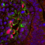
 Lung Specific Gene Regulation and Transcription Factors. Double immunofluorescent staining shows a transcription factor (pink) in the nucleus of endothelial cells (green) of a primitive developing vessel in the fetal mouse lung E13.0.  Nuclei were labeled with DAPI (blue) and the developing epithelium is on the right of the picture. (Picture courtesy of the Le Cras and Whitsett laboratories.)

Vrushank Davé, PhD: Transcriptional/signal networks in lung development and cancer

Machiko Ikegami, MD, PhD: Transcriptional control of surfactant maturation

Anne-Karina Perl, PhD: Lung repair processes and progenitor cells in lung development

John Shannon, PhD: Lung morphogenesis and differentiation, and FGFs

Susan Wert, PhD:  Histopathology of abnormal lung development, injury / repair  and human surfactant dysfunctions / mutations.

Jeffrey Whitsett, MD: Lung transcriptional control, epithelial patterning and differentiation

Kathryn Wikenheiser-Brokamp, MD, PhD: Epithelial cell growth regulation, pathways in lung cancer

Yan Xu, PhD: Bioinformatics, systems biology of lung development and disease

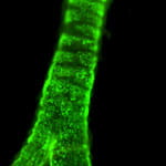
Lung Progenitor / Stem Cells.  Fluorescent staining detects cells, which have originated from a common progenitor source, populating the tracheal rings. (Picture courtesy of the Perl and Whitsett laboratories.)


Timothy Le Cras, PhD: Lung development, pathogenesis of lung remodeling

Anne-Karina Perl, PhD: Lung repair processes and progenitor cells in lung development

John Shannon, PhD: Lung morphogenesis and differentiation, and FGFs

Susan Wert, PhD: Histopathology of abnormal lung development, injury / repair  and human surfactant dysfunctions / mutations.

Jeffrey Whitsett, MD: Lung transcriptional control, epithelial patterning and differentiation

Kathryn Wikenheiser-Brokamp, MD, PhD:  Epithelial cell growth regulation, pathways in lung cancer

Yan Xu, PhD: Bioinformatics, systems biology of lung development and disease

Lung development phases: human and mouse.
Click for larger image.