Bindarit: A Potential Treatment for Protecting Neural Development in Neonatal Hydrocephalus
Published March 2022 | Journal of Neuroscience
The anti-inflammatory agent bindarit may restore cognitive and motor function damaged by neonatal hydrocephalus, investigators at Cincinnati Children’s have found.
Research led by Francesco Mangano, DO, and June Goto, PhD, finds that administering bindarit significantly supports healthy postnatal cerebral cortical development. The findings, based on a mouse model, go beyond what is commonly known about neonatal hydrocephalus—that neuroinflammation and glial cell activation occur as a reaction to pressure.
“It’s not been studied whether it’s also related to cognitive or motor dysfunction in those patients,” Goto says. “We thought we could find a new target to help improve neural cell development and brain function.” Mangano and Goto identified cortical neuropil maturation defects such as:
- Impaired maturation of excitatory synapses
- Dendritic arborization of inhibitory neurons
- Loss of homeostatic microglia
- Hindlimb locomotor defects
“The hydrocephalus mice showed spastic hind limb movements in the swim test,” Goto says. “The bindarit treatment supported both neuronal and glial cell development and improved those movements.”
Hydrocephalus prevents proper myelination and devel-opment of cortical neuronal cells. Bindarit blocks nuclear factor (NF)-kB activation and pro-inflammatory microglial activation. This restores the cortical neuropil thinning and synaptic maturation defects in prh mutant mouse brains.
These study results open the potential for the therapeutic use of anti-inflammatory reagents like bindarit. There may be opportunities to prevent microglial activations or turn off damaging responses, Mangano says. Future studies of bindarit treatment combined with CSF diversion surgery may provide long-term benefits supporting neuronal development in neonatal hydrocephalus.
Myelination and Vascularization in prh White Matter with Bindarit Treatment
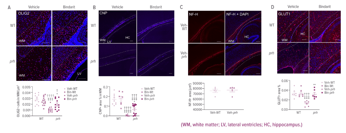
B. The mature myelination marker CNP positive staining area % in WM shows significant reduction of myelination in veh-prh and partial recovery in bin-prh.
C. Neurofilamentlabeled axons (NF-H) positive area (μm2) in WM shows no significant difference between veh-WT and veh-prh.
D. Endothelial cell marker, GLUT1 positive staining area per WM (%) shows reduced vascular densities in veh-prh and significant recovery in bin-prh.


